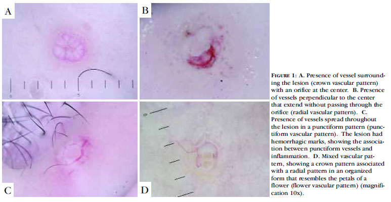
BACKGROUNDS: Although easily diagnosed, molluscum contagiosum may present as a single lesion or as several small, inflamed lesions of difficult diagnosis. OBJECTIVE: To describe the dermoscopic characteristics of molluscum contagiosum and to compare the findings from clinical examination and dermoscopy. METHODS: Histopathologically confirmed lesions were evaluated clinically and dermoscopically in 57 patients. RESULTS: At clinical examination and dermoscopy of 211 lesions, orifices were visualized in 50.24% and 96.68% of the lesions, and vessels in 6.16% and 89.10%, respectively. The vascular patterns found in the 188 lesions in which vessels were found at dermoscopy were the crown (72.34%), radial (54.25%) and punctiform patterns (20.21%). Half of the 188 lesions had a combination of vascular patterns, with the flower pattern (a new vascular pattern) being found in 19.68% of cases. More orifices and vessels were identified at dermoscopy than at clinical examination, including cases with inflammation or perilesional eczema and small lesions. Punctiform vessels were associated with inflammation, excoriation and perilesional eczema. CONCLUSIONS: Dermoscopy performed on molluscum contagiosum lesions proved superior to dermatological examination even in cases in which clinical diagnosis was difficult. The presence of orifices, vessels and specific vascular patterns aids diagnosis, including differential diagnosis with other types of skin lesion.
Dermatology; Diagnostic equipment; Microscopy; Molluscum contagiosum


