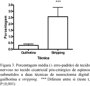
The post-operative healed tissues in horses submitted to two digital neurectomy techniques, the guilhotine (GT) and the stripping (ST), were evaluated by macroscopy and microscopy. Fourteen months after surgery, 32 samples of scar tissue were collected from four mares that had the members experimentally submitted to both surgical techniques. By macroscopy, the dimensions of the scar tissue of the proximal stump and the distance between nerve stumps were taken. By microscopy, the proportion of nervous tissues in the scar tissue was quantified by histomorphometry. There were no differences between the scar tissue dimensions, but the distance between stumps was 5.6-fold greater in ST subjects. Histologically, connective tissue, macrophages, and thin nervous fibers were observed in scar tissue present in animals of both groups. Nodular structures composed by nervous fascicules were visualized in 56.2% (9/16) of the samples collected from the ST group. The mean percentage of the nervous tissue in scar tissue was 0.31% in GT samples and 2.6% in ST samples (P<0.001). After ST, a longer time to the return of the sensibility may occur due to the greater distance between stumps. However, greater proportion of nervous tissue in the scar tissue suggests that the use of this technique favors nervous regeneration.
horse; neurectomy; pain; neuromas; reinervation


