Resumos
O objetivo deste relato de caso é descrever a ocorrência de plasmocitoma em bulbo peniano de um cão, classificado como uma doença extramedular não cutânea de localização rara e casuística inédita. Um cão, sem raça definida, com sete anos de idade e pesando 15kg, não castrado, apresentou histórico clínico de anorexia, vômitos, anúria e constipação. Ao exame específico da genitália externa, foi encontrada uma massa em bulbo peniano durante a inspeção do prepúcio, aderida à pele e encapsulada, extremamente firme e arredondada, medindo cerca de 6cm de diâmetro. Por meio da ultrassonografia dessa estrutura, foi observado aumento do volume regional com ecotextura heterogênea e ecogenicidade mista, além de neovascularização tecidual ao Doppler colorido. Foi realizada biópsia da massa, sendo verificada a presença de neoplasia de células redondas. A caracterização do tumor foi realizada pela imuno-histoquímica, e as células neoplásicas foram imunoexpressas para CD79a e MUM1, indicando o diagnóstico de plasmocitoma extramedular. Embora os tumores penianos em cães sejam os predominantemente venéreos transmissíveis (TVT), e os plasmocitomas sejam neoplasias raras nessa localização, este relato de caso fornece com ineditismo a ocorrência de plasmocitoma extramedular em bulbo peniano de cão, condição ainda não descrita em veterinária.
canino; plasmocitoma; sistema reprodutivo
Non-cutaneous extramedullary plasmacytomas are relatively rare in dogs, affecting mainly the oral cavity and bowel loops. The involvement of the penile bulb has not been described, a fact of great importance for obstetric and veterinary oncology. The aim of this case report is to describe the occurrence of plasmacytoma in a dog's penile bulb, classified as a non-cutaneous extramedullary disease of rare location and unpublished casuistry. A non castrated dog of undefined breed, with seven years of age and weighing 15kg, presented clinical history of anorexia, vomiting, anuria and constipation. By specific examination of the external genitalia, a penile bulb mass was found in the preputial inspection, which was adhered to the encapsulated skin, extremely firm and rounded, measuring approximately 6 cm in diameter. By ultrasound evaluation of the structure in the penile bulb an increase of regional volume with heterogeneous echotexture and mixed echogenicity and tissue neovascularization upon color Doppler was observed. Incisional biopsy of the mass was performed and showed the presence of neoplasia of round cells. The characterization of the tumor was performed by immunohistochemistry and the neoplastic immuno cells were expressed CD79a and MUM1, indicating the diagnosis of extramedullary plasmacytoma. Although the penile tumors in dogs are predominantly transmissible venereal tumors (TVT) and plasmocytomas are rare neoplasms in this location, this case report provides a novel occurrence of extramedullary plasmacytoma in the penile bulb of a dog, a condition not yet described in veterinary.
canine; plasmacytoma; reproductive system
INTRODUCTION
Neoplastic disorders related to plasma cells, described in veterinary, comprise a group of affections with highly variable clinical behavior among which are: multiple myeloma, solitary bone plasmacytoma, extramedullary plasmacytomas (subdivided in cutaneous and non-cutaneous) and, less commonly, leukemia of plasma cells and Waldenstrom's macroglobulinemiade (Jacobs et al. 2002JACOBS, R.M.; MESSICK, J.B.; VALLI, V.E. Tumors of the hemolymphatic system. In: MEUTEN, D.J. (Ed). Tumors in domestic animals. Ames: Iowa State Press, 2002. p.161-164.; Hayes et al. 2007HAYES, A.M.; GREGORY, S.P.; MURPHY, S. et al. Solitary extramedullary plasmacytoma of the canine larynx. J. Small Anim. Pract., v.48, p.288-291, 2007.).
Non-cutaneous extramedullary plasmacytomas are relatively rare in dogs, affecting mainly the oral cavity and bowel loops (Wrigth et al. 2008). Though there is a report described in the literature regarding the occurrence of an extramedullary plasmacytoma in penile glans in dog (Kim et al. 2010KIM, M.S.; KIM, D.H.; CHOI, U.S. Penile extramedullary plasmacytoma in a dog. Reprod Domest Anim, v.45, p.e454-e457, 2010.), the involvement of the penile bulb has not been described, a fact of great importance for obstetric and veterinary oncology.
Considering that the description of plasmacytoma in a dog's penile bulb described in this report may be classified as a non-cutaneous extramedullary disease of rare location, the aim of this case report is to describe the occurrence of neoplastic condition, concerning the main clinical features observed in this unpublished casuistry.
CASUISTRY
A non castrated dog of undefined breed, with seven years of age and weighing 15kg, was attended in Veterinary Hospital Governador Laudo Natel - Unesp Jaboticabal with a clinical history of anorexia, vomiting, anuria (for one day) and constipation (for three days). According to the owner, the animal had been diagnosed seven months previously with chronic kidney disease (stage 2) and reported not having started any type of therapy.
After anamnesis, the animal was subjected to physical examination and 7% dehydration was observed, as well as congested mucous membranes, hypothermia (rectal temperature of 36.3ºC), heart rate of 90 beats per minute, respiratory rate of 20 breaths per minute and normal lymph nodes. From the abdominal palpation, distended bladder and abdominal pain was verified.
During the specific examination of the external genitalia, a penile bulb mass was found in the preputial inspection, which was adhered to the encapsulated skin, extremely firm and rounded, measuring approximately 6 cm in diameter, possibly promoting mechanical obstruction of the urethra, once the owner reported that the dog had been having problems to urinate.
For further information, the blood count, biochemical profile (creatinine, urea, alanine aminotransferase/ALT, alkaline phosphatase/FA, phosphorus and serum albumin), urinalysis, abdominal and regional ultrasonography (preputial structure and bulb), chest x-ray and bulb mass biopsy from fine needle aspiration were performed.
Hematological parameters showed a normocytic anemia, normochromic (erythrocytes 3.3x10⁶/uL), leukocytosis per neutrophils (total leukocytes 20,400/uL). The serum chemistries demonstrated azotemia (creatine 3.6mg/dl; urea: 163mg/dl) hyperphosphataemia (phosphorus 7.92mg/dL); hypoalbuminaemia (albumin 1.2mg/dL); hepatic enzymes within normal measures (ALT: 40IU/L, and F: 41IU/L). The urinalysis showed isosthenuria (density: 1.010) and proteinuria. Cytopathology mass in the penile bulb was suggestive of neoplasia of round cells, and it was not possible to accurately determine the origin, due to the degree of cell differentiation.
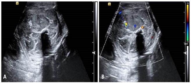
Ultrasound examination was observed to increase the splenic silhouette, showing the presence of heterogeneous echotexture and mixed echogenicity (presence of solid components - diffuse echogenic points) and tissue neovascularization (color Doppler mode); During the ultrasound evaluation of the structure in the penile bulb, an increase in the volume of the preputial region with loss of tissue definition was found, as well as tissue with heterogeneous echotexture and mixed echogenicity (solid and liquid components) and tissue neovascularization on color Doppler mode (Figure 1).
Ultrasound images of structure in the penile bulb region of a dog, to the B-mode (A) and color Doppler (B). Note in (A) the presence of mixed echogenicity and heterogeneous echotexture of the evaluated structure and (B) the occurrence of tissue neovascularization.
To assess the flow and urinary debt, the patient was catheterized and maintained in therapeutic protocol intravenous fluid, antiemetic drugs (ondansetron 0.5mg/kg ranitidine and 2mg/kg every 12 hours, intravenously), nutrition via nasogastric tube and aluminum hydroxide administration associated with feed for correction of hyperphosphatemia.
After stabilization and general improvement of the clinical condition, the animal was submitted to the incisional biopsy of the mass from the penile bulb. The material was sent for histopathological evaluation. After biopsy, analgesia (tramadol 4mg/kg-TID and dipyrone 25mg/kg-TID) and antibiotics (cephalexin 30mg/kg-BID) for 7 days were added to the therapeutic protocol.
Histopathological diagnosis showed the presence of neoplasia of round cells, forming cords along the dermis. The cells had hyperstained nuclei, small and medium, peripherals and moderate amount of eosinophilic granular cytoplasm with rare mitotic figures. Characterization of the tumor was performed by immunohistochemistry. The tissue sections were routinely processed for histology and included in paraffin and were placed over blades previously assigned. Antigen retrieval by moist heat method was carried out in steam cooker, for 20-30min. Incubation with primary antibodies was carried out overnight at 4ºC. To print the image we used the Advance system. The coloration was performed with 3.3 - diaminobenzidine and counterstaining with hematoxylin. External and/or internal controls were used to validate the reaction. For positive controls normal lymph nodes were used, and as a negative control the primary antibody was omitted and replaced it with sodium chloride 0.9%.
The neoplastic cells immuno expressed CD79a (DAKO - JCB 117 - dilution 1:150) and MUM1 (Cell Marque - MRQ.43 - dilution 1:100) (Figure 2); did not express CD3 (canine CD3 SEROTEC - MCA17745 - dilution 1:300), PAX5 (Cell Marque - SP34 - dilution 1:100), Lysozyme (DAKO - AEC32117 - dilution 1:5000) and Tryptase (Cell Marque - G3 - dilution 1:300) (Fig. 3). The interpretation of immunohistochemical examination along with the morphological results indicated the diagnosis of neoplasia of plasma cells (extramedullary plasmacytoma).
Immunohistochemical image of penile bulb tissue of dog with (A) positive membrane-associated staining pattern for CD79a and (B) positive nuclear staining pattern to MUM-1.
Immunohistochemical image with (A) negative staining for CD3 (B) negative staining for PAX5 (C) negative staining for lysozyme (D) negative staining for tryptase.
Due to the worsening of clinical symptoms of the canine patient on the subsequent days, the performance of surgery for mass removal was impossible. Supportive treatment for chronic kidney disease and analgesia were conducted, but there was no satisfactory clinical response and the animal evolved to neurological alterations, side decubitus and stupor, with consequent death.
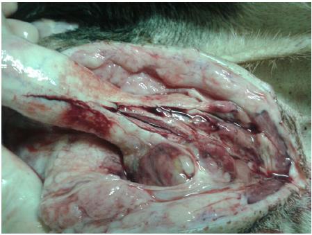
In the post mortem examination, in the macroscopic evaluation the presence of a mass in the penile bulb was confirmed with light coloring and firm appearance, extremely adhered (Figure 4). The histopathological splenic assessment revealed only a focus of benign hyperplasia.
Photography of the structure in the penile bulb region of the canine, performed during necropsy of the animal, observing the presence of an adhered mass, necrotizing and reddened areas.
DISCUSSION
Plasmocytomas extramedullary in dogs are uncommon neoplasms that affect adult and elderly animals, with a mean age of 9.4 years, which corroborates with the animal in the present study (Ramos-Vara et al., 2007RAMOS-VARA, J.A.; MILLER, M.A.; VALLI, V.E.O. Immunohistochemical Detection of Multiple Myeloma 1/Interferon Regulatory Factor 4 (MUM1/IRF-4) in Canine Plasmacytoma: Comparison with CD79a and CD20. Vet. Pathol., v.44, p.875-884, 2007.). The main sites of involvement of extramedullary plasmacytomas include the skin, responsible for 86% of cases, followed by oral mucosa and lips (9%), colon and rectum (4%) and other sites such as the stomach, spleen, liver, uterus, eye, larynx, trachea, genitalia, central nervous system and nasal cavity corresponding to only 1% of the total cases (Vail et al., 2012VAIL, D.M.; PINKERTON, M.E.; YOUNG, K.M. Hematopoietic tumours. In: WITHROW, S.J.; VAIL, D.M.; PAGE, R.L. (Eds). Small animal clinical oncology. 5.ed. St Louis: Elsevier, 2012. p.665-679.). In addition, Ramos-Vara et al. (2007) observed in 109 cases of extramedullary plasmacytoma in dogs, the occurrence of skin lesions in 80 patients, followed by 18 cases in the oral region, 5 patients with intestinal alteration (rectum) and 2 dogs with splenic involvement. Other affected locations were the liver, lung and brain, but the occurrence of neoplasia in the penile region had never been observed, a fact that denotes great relevance to the present case report.
The main tumors that involve the penile region of dogs are transmissible venereal tumors (TVT) and squamous cell carcinomas, but there are other histological types such as adenomas, papillomas, lymphoma, mast cell tumors, hemangiomas, melanoma, fibrosarcoma and osteosarcoma of the penis bone (MacLachlan and Kennedy, 2002MACLACHLAN, N.J.; KENNEDY, P.C. Tumors of the genital systems. In: MEUTEN, D.J. (Ed). Tumors in domestic animals. Ames: Iowa State Press, 2002. p.570-573.). Although there is a description, in the veterinary literature, of a case of plasmacytoma in a canine penis, it was located in the penis glans and appeared as a single nodule of approximately 1.3cm (Kim et al., 2010), unlike the case reported where the mass was located in the penile bulb and presented invasive behavior. This is the first report describing the occurrence of plasmacytoma in a dog's penile bulb.
Considering that the patient in this report had access to the street and was not castrated, by the location of the mass the first differential diagnosis was transmissible venereal tumor (TVT). However, the firm consistency presented by the mass, the presence of a capsule and the degree of adhesion site were not compatible with TVT, since this disease usually presents itself as a friable mass, ill-defined with the presence of site hemorrhage (Das and Das, 2000DAS, U.; DAS, A.K. Review of canine transmissible venereal sarcoma. Vet Res Commun, v.24, p.545-556, 2000.).
The cytopathology of the neoplasia of undifferentiated round cell didn't allow the completion of the diagnosis, since both TVT as plasmacytoma belong to the group of round cell tumors. The TVT may still be subdivided by cytological analysis in two different ways: linfocitoide (round morphology, scant cytoplasm and rounded nucleus) and plasmacytoid (this appears as an ovoid morphology, abundant cytoplasm, eccentric nucleus and a larger number of vacuoles), being that the venereal tumors with lesser vacuoles, especially linfocitoide are differential diagnosis of other round cell neoplasms, including plasmacytomas, as well as the plasmacytoid with scarce vacuolization that can be confused with other round cell tumors such as plasmacytoma (Flores et al., 2012FLORES, M.M.; PEDRAZA, F.; GRANDI, F. et al. Cytologic subtypes of canine transmissible venereal tumor. Vet. Clin. Pathol., v.41, p.4-5, 2012.; Rocha et al., 2014ROCHA, N.S.; TREMORIL, T.M.; CARNEIRO, J.A.M. Fine needle aspiration cytology in the diagnosis of canine cutaneous transmissible venereal tumor - case report. Open J. Vet. Med., v.4, p.204-209, 2014.), which can hinder the diagnosis in a cytological preparation of which characteristics are not as well differentiated as in the cytological examination of the animal in question.
The ultrasonographic results allowed for a better clinical characterization of the patient (by the exclusion of metastasis area on chest radiography) and mass in the penile bulb. Nodules presenting heterogeneous sonographic findings (echotexture and echogenicity), as observed in the present report, and neovascularization, are suggestive of neoplastic masses (Feliciano et al., 2014FELICIANO, M.A.R.; MARONEZI, M.C.; PAVAN, L. et al. ARFI elastography as a complementary diagnostic method for mammary neoplasia in female dogs - preliminary results. J. Small Anim. Pract., v.55, p.504-508, 2014.; Assis et al., 2015ASSIS, A.R.; GARCIA, D.A.A.; FELICIANO, M.A.R. Sistema Reprodutor Masculino. In: FELICIANO, M.A.R.; CANOLA, J.C.; VICENTE, W.R.R. (Eds). Diagnóstico por imagem em cães e gatos. São Paulo: Editora MedVet, 2015. p.368-409.).
The histopathological evaluation associated with immunohistochemical examination was paramount for the diagnostic conclusion of this case. Positive immunostaining for CD79a and MUM-1 (IRF4) is important in the characterization of plasmacytomas. Ramos-Vara et al. (2007) evaluated the immunostaining of CD79a and MUM-1 (IRF4) in 109 cases of plasmacytomas in dogs and positive immunostaining for MUM-1 (IRF4) and CD79a was respectively for 93.5% and 56.2% of the cases of plasmacytoma. The MUM1 marker (IRF4) proved to be more specific and sensitive than the CD79a, but the combined assessment of both markers presents a sensitivity of 95.5% in the diagnosis of plasmacytoma in dogs.
An important difference in this case is diffuse lymphomas of large B cell, but only 21.2% of B-cell lymphomas in this study expressed MUM-1 (IRF4), which together with the morphological results allowed a precise distinction. These data were used in this study for the differentiation between lymphoma B and extramedullary plasmacytoma (Varas-Ramos et al., 2007).
Cutaneous solitary plasmacytomas and mucous usually show favorable prognosis and the surgical resection is already curative. In a large-scale evaluation of cases of cutaneous and mucosal plasmacytoma, the local recurrence rate was 5% and 2% of metastasis in a lymph node (Cangul et al., 2002CANGUL, I.T.; WUNEN, M.; VAN GARDEREN, E. et al. Clinico-pathological aspects of canine cutaneous and mucocutaneous plasmacytomas. J. Vet. Med. A Physiol. Pathol. Clin. Med., v.49, p.307-312, 2002.). Dogs with extramedullary plasmacytoma in the gastrointestinal tract or other organs of the abdominal cavity treated with surgery associated or not to chemotherapy may have long-term survival (Jacobs et al., 2002; Vail et al., 2012). Surgical intervention in this case could lead to a good local control of the disease, but the same was not possible due to the overall clinical condition of the animal as a result of chronic kidney disease that was in progress and was probably responsible for the unfavorable evolution of the case.
In dogs there are few reports of progression of extramedullary plasmacytomas for multiple myeloma, although this condition has been described (Vail et al., 2012). In this case the progression to multiple myeloma cannot be assessed due to the rapid evolution of the case.
CONCLUSION
Although the penile tumors in dogs are predominantly transmissible venereal tumors (TVT) and plasmocytomas are rare neoplasms in this location, they should be included in the differential diagnosis of nodular alterations of the reproductive tract of canine, especially in cases where the cytological evaluation of round cell tumors is not confirmatory regarding to the precise origin of the tumor.
- ASSIS, A.R.; GARCIA, D.A.A.; FELICIANO, M.A.R. Sistema Reprodutor Masculino. In: FELICIANO, M.A.R.; CANOLA, J.C.; VICENTE, W.R.R. (Eds). Diagnóstico por imagem em cães e gatos. São Paulo: Editora MedVet, 2015. p.368-409.
- CANGUL, I.T.; WUNEN, M.; VAN GARDEREN, E. et al. Clinico-pathological aspects of canine cutaneous and mucocutaneous plasmacytomas. J. Vet. Med. A Physiol. Pathol. Clin. Med., v.49, p.307-312, 2002.
- DAS, U.; DAS, A.K. Review of canine transmissible venereal sarcoma. Vet Res Commun, v.24, p.545-556, 2000.
- FELICIANO, M.A.R.; MARONEZI, M.C.; PAVAN, L. et al. ARFI elastography as a complementary diagnostic method for mammary neoplasia in female dogs - preliminary results. J. Small Anim. Pract., v.55, p.504-508, 2014.
- FLORES, M.M.; PEDRAZA, F.; GRANDI, F. et al. Cytologic subtypes of canine transmissible venereal tumor. Vet. Clin. Pathol., v.41, p.4-5, 2012.
- JACOBS, R.M.; MESSICK, J.B.; VALLI, V.E. Tumors of the hemolymphatic system. In: MEUTEN, D.J. (Ed). Tumors in domestic animals. Ames: Iowa State Press, 2002. p.161-164.
- HAYES, A.M.; GREGORY, S.P.; MURPHY, S. et al. Solitary extramedullary plasmacytoma of the canine larynx. J. Small Anim. Pract., v.48, p.288-291, 2007.
- KIM, M.S.; KIM, D.H.; CHOI, U.S. Penile extramedullary plasmacytoma in a dog. Reprod Domest Anim, v.45, p.e454-e457, 2010.
- MACLACHLAN, N.J.; KENNEDY, P.C. Tumors of the genital systems. In: MEUTEN, D.J. (Ed). Tumors in domestic animals. Ames: Iowa State Press, 2002. p.570-573.
- RAMOS-VARA, J.A.; MILLER, M.A.; VALLI, V.E.O. Immunohistochemical Detection of Multiple Myeloma 1/Interferon Regulatory Factor 4 (MUM1/IRF-4) in Canine Plasmacytoma: Comparison with CD79a and CD20. Vet. Pathol., v.44, p.875-884, 2007.
- ROCHA, N.S.; TREMORIL, T.M.; CARNEIRO, J.A.M. Fine needle aspiration cytology in the diagnosis of canine cutaneous transmissible venereal tumor - case report. Open J. Vet. Med., v.4, p.204-209, 2014.
- VAIL, D.M.; PINKERTON, M.E.; YOUNG, K.M. Hematopoietic tumours. In: WITHROW, S.J.; VAIL, D.M.; PAGE, R.L. (Eds). Small animal clinical oncology. 5.ed. St Louis: Elsevier, 2012. p.665-679.
- WRIGHT, Z.M.; ROGERS, K.S.; MANSELL, J. Survival data for canine oral extramedullary plasmacytomas: a retrospective analysis (1996-2006). J. Am. Anim. Hosp. Assoc., v.44, p.75-81, 2008.
Datas de Publicação
-
Publicação nesta coleção
Mar-Apr 2016
Histórico
-
Recebido
28 Abr 2015 -
Aceito
15 Nov 2015