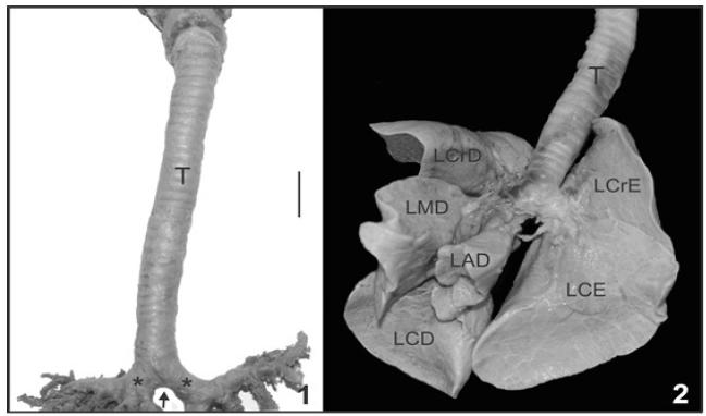
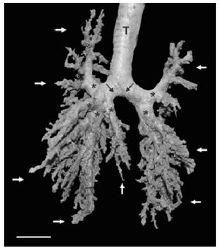
No characteristic of living beings is as primal as breathing, and the lungs are the main organs in the respiratory system. This study aims to describe the macroscopic aspects of the trachea, bronchus and lung lobes and microscopic aspects of the bronchi of raccoon lungs and compare with data from the literature studies performed with wild and domestic mammals. We used three samples of Procyon cancrivorus, which were fixed in aqueous 10% formaldehyde, lungs and trachea were dissected and photographed with a digital camera (Sony a200 Camera, 10.2mpx). For the identification of microscopic characteristics, fragments were collected from each bronchus following routine histological techniques. The Procyon cancrivorus lung is divided into four lobes, with two right and left lobes. The trachea has about 31-34 rings. The extrapulmonary bronchi divides into left and right, where the right is divided into lobar bronchi cranial, middle, accessory and caudal lobes and the left in cranial and caudal, with their respective segmental bronchi. Microscopically the bronchial epithelium has prismatic pseudo-ciliated and goblet cells with bundles of smooth muscle fibers, plates of hyaline cartilage and elastic fibers. Knowledge of the morphology of these organs in wild species helps us in descriptive studies and / or comparisons between species.
Anatomy; bronchi; raccoon; trachea


