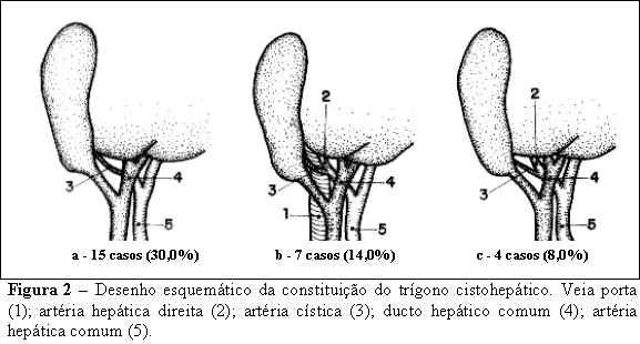
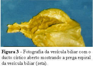
OBJECTIVE: study the morphology of the extrahepatic biliary system and cystichepatic triangle (Calot’s triangle) analysing the disposition, variations and malformations. METHODS: 50 adult cadavers were studied. RESULTS: in 47 cases (94%), the cysticohepatic junction ocurred near the hepatic hilum. In 3 cases (6%), the junction between cystic duct and common hepatic duct ocurred at the level of the hepatopancreatic ampulla (Vater's ampulla). The angle formed by the junction of the cystic duct with the common hepatic duct was less than 30º in 72,4% of the cases. In 23,0%, this angle was between 30 and 45º. In 21,3% was between 45 and 60º. In 2,3% of the cases it was greater than 60º. The common hepatic duct recieved the cystic duct on its right side in the majority of the cases (59,6%); anteriorly in 17% of the cases; posteriorly in 12,8% and on the left side in 10,6% of the cases. In relation to the cystichepatic triangle components the cystic artery was present in 56% of the cases; the portal vein in 36%; the right hepatic artery in 34%; the left hepatic artery in 2% and the proper hepatic artery in 2% of the cases. The dimensions of the cystic duct were 2,53±1,19cm of length and 0,29±0,12cm of diameter. The spiral fold (Heister's valve) was observed in 80% of the cases. The infundibulum of the gallbladder (Hartmann's pouch) was present in 74% of the cases. This knowledge contribute to the practice of video-assisted laparoscopy in this region. CONCLUSION: in cystichepatic triangle the cystic artery was found most frequently.
Extrahepatic biliary system; Video-assisted laparoscopy; Anatomy


