Abstracts
PURPOSE: To evaluate tympanic bulla healing after experimental lateral osteotomy in cats. METHODS: Twenty adult cats were submitted to unilateral lateral bulla osteotomy and divided into two groups: cats of B1 group (n=10) were euthanized at 8 weeks and cats of B2 group (n=10), at 16 weeks postoperative. RESULTS: Oblique lateral radiographs taken immediately postoperative showed interruption in the contour of the external acoustic meatus of the operated bullae in all cats of both groups (McNemar test: p=0.0010*). This feature was still observed in the radiographs taken after 8 and 16 weeks postoperative (McNemar test: B1 p=0.0020*; B2 p=0.0312*). Macroscopic examination showed that the operated bullae were similar to the normal ones, with preservation of the tympanic cavity. Connective tissue at the osteotomy site was significantly found in the operated bullae in both groups (McNemar test: B1 p=0.0020*; B2 p=0.0010*). The length of connective tissue at the osteotomy site was measured by histomorphometry. There was no statistically significant difference between the values of B1 group and B2 group (Mann-Whitney test: p=0.0524). CONCLUSIONS: Experimental lateral osteotomy did not alter significantly the tympanic bulla conformation and complete regeneration of the tympanic bulla frequently did not occur before 16 weeks of postoperative period.
Ear, middle; Ear neoplasms; Cats
OBJETIVO: Avaliar a morfologia da bulla tympanica de gatos após osteotomia lateral unilateral. MÉTODOS: Foram utilizados 20 gatos distribuídos em dois grupos de 10 animais cada, de acordo com o período de observação: B1 (8 semanas) e B2 (16 semanas). RESULTADOS: Nas radiografias em projeção lateral oblíqua realizadas no pós-operatório imediato observou-se a interrupção do meato acústico externo da bulla tympanica operada de todos os gatos de ambos os grupos (McNemar, p=0,0010*). Esta característica ainda pode ser observada nas radiografias realizadas 8 ou 16 semanas após a cirurgia (McNemar: B1 p=0.0020*; B2 p=0.0312*). Os exames macroscópicos revelaram que a bulla tympanica operada apresentava conformação semelhante a da bulla tympanica normal, com preservação da cavidade timpânica. Na maioria das bullae tympanicae operadas observou-se a presença de tecido conjuntivo na área de osteotomia. O resultado do teste de McNemar foi significante em ambos os grupos (B1 p=0,0020*; B2, p=0,0010*). A histomorfometria mediu a extensão de tecido conjuntivo presente no local da osteotomia. Não houve diferença estatisticamente significante entre os dois grupos (Mann-Whitney, p=0,0524). CONCLUSÕES: A osteotomia lateral não alterou de maneira significativa a conformação da bulla tympanica. A regeneração total da bulla tympanica geralmente não ocorreu antes de 16 semanas de pós-operatório.
Ouvido médio; Neoplasias do ouvido; Gatos
ORIGINAL ARTICLE
SURGICAL SKILLS
Morphological aspects of tympanic bulla after lateral osteotomy in cats1 1 Research performed at Department of Clinics, Surgery and Animal Reproduction, São Paulo State University (UNESP). Araçatuba, Brazil.
Aspectos morfológicos da bulla tympanica de gatos após osteotomia lateral
Adelina Maria da SilvaI; Wilson Machado de SouzaII; Roberto Gameiro de CarvalhoIII; Gisele Fabrino MachadoI; Silvia Helena Venturoli PerriIV
IPhD, Assistant Professor, Department of Clinics, Surgery and Animal Reproduction, UNESP. Araçatuba, Brazil
IIPhD, Full Professor, Department of Biosciences, Production and Animal Health, UNESP. Araçatuba, Brazil
IIIMaster, Assistant Professor, Department of Biosciences, Production and Animal Health, UNESP. Araçatuba, Brazil
IVPhD, Assistant Professor, Department of Biosciences, Production and Animal Health, UNESP. Araçatuba, Brazil
Correspondence Correspondence: Adelina Maria da Silva São Paulo State University (UNESP). Araçatuba, Brazil Department of Clinics, Surgery and Animal Reproduction Rua Clóvis Pestana, 793 16050-680 Araçatuba SP Brazil adelinasilva@uol.com.br
ABSTRACT
PURPOSE: To evaluate tympanic bulla healing after experimental lateral osteotomy in cats.
METHODS: Twenty adult cats were submitted to unilateral lateral bulla osteotomy and divided into two groups: cats of B1 group (n=10) were euthanized at 8 weeks and cats of B2 group (n=10), at 16 weeks postoperative.
RESULTS: Oblique lateral radiographs taken immediately postoperative showed interruption in the contour of the external acoustic meatus of the operated bullae in all cats of both groups (McNemar test: p=0.0010*). This feature was still observed in the radiographs taken after 8 and 16 weeks postoperative (McNemar test: B1 p=0.0020*; B2 p=0.0312*). Macroscopic examination showed that the operated bullae were similar to the normal ones, with preservation of the tympanic cavity. Connective tissue at the osteotomy site was significantly found in the operated bullae in both groups (McNemar test: B1 p=0.0020*; B2 p=0.0010*). The length of connective tissue at the osteotomy site was measured by histomorphometry. There was no statistically significant difference between the values of B1 group and B2 group (Mann-Whitney test: p=0.0524).
CONCLUSIONS: Experimental lateral osteotomy did not alter significantly the tympanic bulla conformation and complete regeneration of the tympanic bulla frequently did not occur before 16 weeks of postoperative period.
Key words: Ear, middle. Ear neoplasms. Cats.
RESUMO
OBJETIVO: Avaliar a morfologia da bulla tympanica de gatos após osteotomia lateral unilateral.
MÉTODOS: Foram utilizados 20 gatos distribuídos em dois grupos de 10 animais cada, de acordo com o período de observação: B1 (8 semanas) e B2 (16 semanas).
RESULTADOS: Nas radiografias em projeção lateral oblíqua realizadas no pós-operatório imediato observou-se a interrupção do meato acústico externo da bulla tympanica operada de todos os gatos de ambos os grupos (McNemar, p=0,0010*). Esta característica ainda pode ser observada nas radiografias realizadas 8 ou 16 semanas após a cirurgia (McNemar: B1 p=0.0020*; B2 p=0.0312*). Os exames macroscópicos revelaram que a bulla tympanica operada apresentava conformação semelhante a da bulla tympanica normal, com preservação da cavidade timpânica. Na maioria das bullae tympanicae operadas observou-se a presença de tecido conjuntivo na área de osteotomia. O resultado do teste de McNemar foi significante em ambos os grupos (B1 p=0,0020*; B2, p=0,0010*). A histomorfometria mediu a extensão de tecido conjuntivo presente no local da osteotomia. Não houve diferença estatisticamente significante entre os dois grupos (Mann-Whitney, p=0,0524).
CONCLUSÕES: A osteotomia lateral não alterou de maneira significativa a conformação da bulla tympanica. A regeneração total da bulla tympanica geralmente não ocorreu antes de 16 semanas de pós-operatório.
Descritores: Ouvido médio. Neoplasias do ouvido. Gatos.
Introduction
Total ear canal ablation with lateral tympanic bulla osteotomy (TECA/LBO) is most frequently indicated for cats with neoplastic disease of the ear.1-3 The most common ear tumors in these animals are ceruminous gland adenocarcinoma and squamous cell carcinoma. 1-7 The tympanic bulla in cats is divided into a dorsolateral and a ventromedial compartment by an incomplete septum in the caudomedial aspect of the smaller compartment that allows communication between the cavities. Thus, when lateral bulla osteotomy is performed, part of this septum must be removed for proper curettage and drainage of both compartments, and the surgeon has to avoid injuries to the structures of hearing and balance or to the nerve tracts that cross the middle ear. 2,8
A better understanding of the consequences of TECA/LBO may improve the management of recurrences, reoperations and changes in middle-ear function, since this procedure is increasingly common in cats. In spite of this, we have not found experimental studies about this surgery in cats. In fact, healing of tympanic bulla after TECA/LBO has been evaluated mostly in dogs, whose bulla consists of a single compartment.9,10 Therefore, this study evaluates the morphology of the tympanic bulla after TECA/LBO in cats by radiographic, macroscopic, histopathological and histomorphometric examinations. Risks of nerve and inner ear damage were also assessed.
Methods
This study was approved by the Ethics Committee for Animal Research of the São Paulo State University campus Araçatuba.
Animals and experimental design
Twenty healthy young adult (aging between 8 and 24 months) mixed breed cats (weighing 3,0 ± 0,4kg) were utilized. Ten spayed females and 10 neutered males were used. Experimental total ear canal ablation with lateral tympanic bulla osteotomy (Figure 1) was performed at random in either the right or left ear. Animals were divided into two groups, according to the postoperative observation time: cats of B1 group (n=10) were euthanized at 8 weeks and cats of B2 group (n=10), at 16 weeks postoperative.
Surgical procedure
Food was withheld for 12 hours prior to anesthesia. The cats were premedicated with atropine (0.05mg/kg SC) and xylazine (2mg/kg SC). Anesthesia was induced with ketamine (15mg/kg, SC), and maintained with thiopental (5mg/kg IV) and fentanil (0.0125mg/kg IM). Lactated Ringer's solution (10ml/kg IV) was administered throughout anesthesia. Enrofloxacin (5mg/kg SC) and ketoprofen (2mg/kg SC) were administered preoperatively and for 3 days postoperatively.
The lateral cranial and cervical regions were clipped, prepared, and draped under sterile technique. Cats were positioned in lateral recumbency. A T-shaped skin incision was made over the vertical ear canal, encircling the external ear canal opening. Using blunt and sharp dissection, the cartilage of the vertical ear canal was dissected from the overlying skin and surrounding soft tissue. Upon reaching the osseous external acoustic meatus, the cartilaginous ear canal was transected with scissors. A rongeur was used to remove the ventral rim of the osseous external acoustic meatus and a portion of the lateral wall of the tympanic bulla (circa 8mm x 5mm). Removal of the tympanic membrane and malleus was attempted with a curved mosquito hemostat. The septum of the bulla was opened with a curette. The epithelium and lamina propria lining the osseous external acoustic meatus was removed with an electric driven burr. The tympanic cavities of the two compartments were curetted and irrigated with 0.9% saline. A 1-cm diameter Penrose drain was placed at the edge of the tympanic cavity, exited through a separate stab incision in the skin ventrally to the surgical wound. Subcutaneous layer was closed with 4-0 polyglactin 910. Skin closure was with 4-0 nylon. The Penrose drain was removed after 3 days and skin sutures removed on day 7.
Postoperative observations and radiographs
After bulla osteotomy the cats were monitored for clinical signs of Horner's syndrome, damage to the inner ear and facial nerve paralysis.
Radiographs were performed immediately after surgery and at the end of the study, i.e., after 8 (B1) and 16 (B2) weeks postoperative. Oblique lateral, ventrodorsal, and open-mouth views of the tympanic bullae were taken.
Macroscopic analysis, histolopathology and histomorphometry
The cats were euthanized with thiopental (45mg/kg IV) after 8 (B1 group) and 16 weeks (B2 group) postoperative. The normal and operated tympanic bullae of each cat obtained at necropsy were fixed in 10% formalin and decalcified in an aqueous solution of 5.66 formalin 10% to 1 of nitric acid (v/v). Then, each of these bullae was divided into two segments longitudinal to the larger axis of the osteotomy. The specimens were processed routinely and thin sections were made and stained with hematoxylin-eosin for microscopic evaluation.
For the histomorphometry, the images of sections were captured using a Leica DMLS microscope connected to a Sony Power HAD 3 CCD video camera, and transferred to an image analyzer (Leica QWIN). In this planimetric study, the connective tissue length at the osteotomy site was measured in mm. The largest value obtained from each cat was included in data analysis.
Statistical analysis
Non-parametric tests were utilized for the data analysis, taking into consideration the nature of the variables. McNemar's test was used to compare data between operated bullae and contralateral unoperated bullae. Mann-Whitney's test was used to compare histomorphometric data between groups B1 and B2. Significance was set at p£0.05.
Results
None of the operated cats showed clinical signs of either Horner's syndrome or damage to the inner ear. Clinical signs of facial nerve damage were observed in 6 cats. There was one case of facial neuropraxia in one cat from B1 group. The neurological deficit of this animal was resolved by 32 days postoperative. Facial paralysis occurred in 2 cats from B1 group, which remained with a wide palpebral fissure. Postoperative facial neuropraxia was observed in 3 cats from B2 group, but the signs resolved within 25, 27 and 60 days, respectively, for each animal.
Oblique lateral radiographs performed immediately postoperative showed an interruption in the contour of the osseous external acoustic meatus of the operated bulla in all cats of both groups (Figure 2; McNemar's test p=0.0010*) . This feature was still observed in 9 cats from B1 group (p=0.0020*) and in 5 cats from B2 group (p=0.0312*) in the radiographs taken after 8 (Figure 3) and 16 weeks postoperative, respectively.
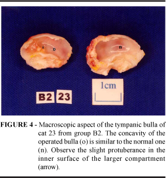
The macroscopic examination revealed that the operated tympanic bullae were similar to the normal ones, with preservation of the tympanic cavity, in both B1 and B2 groups (Figure 4). In group B1, 3 operated bullae presented a slight protuberance in the inner surface of the larger compartment. Also in group B1 the orifice made in the bone septum was still open in one cat, the malleus was still present in 7 tympanic bullae and accumulation of secretion near the external acoustic meatus was observed in 2 cats. In B2 cats the slight protuberance in the inner surface of the larger compartment was present in 3 tympanic bullae. The bone septum was thicker in one cat. The malleus was still present in 4 tympanic bullae. Accumulation of secretion near the external acoustic meatus was observed in 7 cats.
Connective tissue was detected in 9 and 10 operated bullae, from groups B1 and B2, respectively (Figure 5), and only one cat from B1 group showed complete regeneration of the larger compartment after lateral osteotomy. The result of McNemar's test was significant for both groups B1 (p=0.0020*) and B2 (p=0.0010*).
In group B1, the operated tympanic bullae showed a thickened osseous wall in most of the histological sections; thickening of the bone septum also occurred. Ceruminous gland adenitis as well as foci of neutrophils, neovascularization and foamy macrophages in the areas of mononuclear infiltration were observed in the histological sections of one cat of B1 group. The operated tympanic bullae of the B2 group also showed a thickened osseous wall in most of the histological sections. Thickening of the bone septum was also observed. Sebaceous glands, most of them hypertrophied, were found in the histological sections of 6 cats of this group. In many sections the connective tissue was covered by a stratified keratinized squamous epithelium. Histological analysis revealed that the smaller compartment was lined with ciliated pseudostratified columnar epithelium with numerous goblet cells. Three cats from group B2 showed inflammatory processes. Intense infiltration of neutrophils and macrophages with connective tissue proliferation in the lamina propria was found in one cat. Mononuclear infiltrations in the lamina propria and purulent exudate covering the epithelial surface of the tympanic cavity was observed in another cat from B2 group. Severe suppurative inflammation in the osteotomy area filled with connective tissue occurred in the operated tympanic bulla of a third cat from this group. The length of connective tissue at the osteotomy site was measured by the histomorphometry. There was no statistically significant difference between the values for groups B1 and B2 (Mann-Whitney's test, p=0.0524; Table 1) .
Discussion
Retrospective clinical reports cal reports have indicated a prevalence of 12 to 26% of Horner's syndrome after TECA/LBO.1-3 Horner's syndrome, classically presenting with miosis, prolapse of the third eyelid and ptosis of the eyelid, is a result of direct damage to the postganglionic sympathetic fibers coursing to the eyes and eyelids where they pass through the tympanic cavity.8,11-13 No cats in our study showed clinical signs of Horner's syndrome, possibly because during surgery the inner septum was carefully perforated but not removed.
Clinical signs (nystagmus, ataxia and head tilt) indicative of damage to inner ear structures were not observed in any of the 20 cats studied, probably because curettage of the promontory was avoided. Retrospective clinical reports of lateral bulla osteotomy have not made mention of the prevalence of inner ear damage. 1-3
The facial nerve was easily identified coursing along the caudolateral surface of the cartilaginous external acoustic meatus. It was stretched during dissection and facial nerve damage was observed in 6 cats. The clinical signs observed were lack of palpebral reflex and wide palpebral fissure. Drooping of the lip is a clinical sign cited by the literature but it was not observed in our experiment.1,14,15 In the clinical report of 15 cats submitted to TECA/LBO, Williams and White1 verified 4 cases of facial neuropraxia and 7 cases of facial paralysis. In this group, 11 cats had ear neoplastic disease, 3 had polyps and one showed chronic middle ear otitis.
No abnormalities were found in the ventrodorsal radiographs of operated and normal bullae in the immediate postoperative period and at the end of the study in each group (B1 and B2). These abnormalities were probably masked by the overlap of cranial bones as described in a previous report on dogs submitted to experimental unilateral TECA/LBO.10 On the other hand, the open-mouth and oblique lateral radiographs afforded better visualization of the tympanic bulla, detached from the other cranial bones. This allowed the detection of some features in these projections. In all 20 cats from groups B1 and B2, the lateral osteotomy could be consistently detected through the interruption in the contour of the osseous external acoustic meatus in the oblique lateral radiographs taken immediately after surgery. Eight and 16 weeks after the lateral osteotomy, this interruption could still be seen on the radiographs of 9 cats from group B1 and 5 from group B2. Similar results were found in dogs submitted to experimental TECA/LBO.10 Bulla radiography is often useful in evaluating middle-ear disease. Radiographic evidence of bulla disease includes opacities in the tympanic cavity and thickening of the bulla wall.2,3 In the present study, increased density of the tympanic cavity was not found in any of the radiographs performed 8 (B1group) or 16 (B2 group) weeks after lateral osteotomy, indicating that the operated bulla remained filled with air and did not contain tissues or secretions. Thickening of the wall of the bulla was not observed either. A previous report in dogs found similar results.10
The macroscopic examination revealed that the concavity of the operated bullae was similar to the normal ones, with preservation of the tympanic cavity. Only a slight protuberance in the inner surface of the larger compartment was verified in 6 operated bullae. This disagrees with the results of a previous experimental study in dogs, in which the entire lateral wall of the tympanic bulla was removed.9 These authors observed that partial or complete obliteration of the tympanic cavity occurred in most of the dogs due to connective tissue and bone proliferation. In another experimental study in dogs, the bulla osteotomy was restricted to a small area of the lateral wall and the tympanic cavity was preserved almost at its normal volume.10 The bone septum orifice was closed in most of the tympanic bullae of both B1 and B2 groups. However, the communication between the two compartments of the middle ear, naturally present in the caudomedial aspect of the smaller compartment, near the coclear window, was not affected by the osseous proliferation because this occurred only at the osteotomy site in both groups.
Removal of the tympanic membrane and malleus was attempted with a curved mosquito hemostat, but as shown in the macroscopic examination, this procedure was not always successful since the malleus was still present in 11 of the 20 tympanic bullae submitted to lateral osteotomy. No great effort was made to grasp the malleus because this maneuver could push the auditory ossicles against the vestibular window, and consequently cause inner ear damage. Regeneration of the tympanic membrane was not observed in any of the 20 cats. In a experimental study, retention of the tympanic membrane was observed in 2 of 12 dogs submitted to TECA/LBO.9 Removal of the tympanic membrane and malleus was attempted in our study so thatthe epithelium and lamina propria lining the external acoustic meatus could be thoroughly removed.9,14,15 Nevertheless this goal was not totally attained, because the presence of secretion was verified in 9 of the 20 tympanic bullae submitted to lateral osteotomy. As the epithelium and lamina propria lining the external acoustic meatus were not entirely removed with the electric-driven burr, remnantsof sebaceous glands developed postoperatively and secretions of these glands accumulated at the surgical site. That indicates that the curettage of the external acoustic meatus must be carefully done. The performance of this procedure under magnification with a loupe or operating microscope could obtain better results.
The histological examination revealed that only one cat from group B1 showed a tympanic bulla similar to the normal one. In the other 19 cats, connective tissue was observed at the osteotomy site. The operated tympanic bullae of both groups showed a thickened osseous wall and inner septum. Sebaceous glands were found in the histological sections of 6 cats of group B2. Sections of one cat from group B1 and of 3 cats from group B2 presented inflammatory process. There was a trend for the connective tissue length at the osteotomy site to be longer in group B2 than in group B1. The lateral bulla osteotomy was initiated in the ventral rim of the osseous external acoustic meatus. The connective tissue filled the osteotomy area and advanced in a proximal direction, since in this region there was no demarcation by osseous tissue. Furthermore there was an empty space left by the total ear canal ablation.
As a concluding remark, lateral osteotomy did not alter the tympanic bulla conformation and complete regeneration of tympanic bulla frequently did not occur before 16 weeks of postoperative period.
Received: August 29, 2007
Review: October 25, 2007
Accepted: November 27, 2007
Conflict of interest: none
Financial source: FAPESP
- 1. Williams JM, White RAS. Total ear canal ablation combined with lateral bulla osteotomy in the cat. J Small Anim Pract. 1992;33:225-7.
- 2. Trevor PB, Martin RA. Tympanic bulla osteotomy for treatment of middle-ear disease in cats: 19 cases (1984 -1991). J Am Vet Med Assoc. 1993;202:123-8.
- 3. Marino DJ, MacDonald JM, Matthiesen DT, Patnaik AK. Results of surgery in cats with ceruminous gland adenocarcinoma. J Am Anim Hosp Assoc. 1994;30:54-8.
- 4. Legendre AM, Krahwinkel DJ. Feline ear tumors. J Am Anim Hosp Assoc. 1981;17:1035-7.
- 5. Fiorito DA. Oral and peripheral vestibular signs in a cat with squamous cell carcinoma. J Am Vet Med Assoc. 1986;188:71-2.
- 6. Lane IF, Hall DG. Adenocarcinoma of the middle ear with osteolysis of the tympanic bulla in a cat. J Am Vet Med Assoc. 1992;201:463-5.
- 7. Moisan PG, Watson GL. Ceruminous gland tumors in dogs and cats: a review of 124 cases. J Am Anim Hosp Assoc. 1996;32:448-52.
- 8. Little CJL, Lane JG. The surgical anatomy of the feline bulla tympanica. J Small Anim Pract. 1986;27:371-8.
- 9. Mcanulty JF, Hattel A, Harvey CE. Wound healing and brain stem auditory evoked potentials after experimental total ear canal ablation with lateral tympanic bulla osteotomy in dogs. Vet Surg. 1995;24:1-8.
- 10. Silva AM, Fagundes DJ, Taha MO, Goldenberg S, Machado GF, Juliano Y, Novo NF. Aspectos morfológicos da bulla tympanica de cães após osteotomia restrita ventral e lateral. Acta Cir Bras. 2001;16:243-50.
- 11. Barlow CM, Root WS. The ocular sympathetic path between the superior cervical ganglion and the orbit in the cat. J Comp Neurol. 1949;90:195-207.
- 12. Greene GE, Oliver Jr JE. Neurologic examination. In Ettinger SJ. Textbook of veterinary internal medicine. 2ed. Philadelphia: Saunders; 1983. p 419-60.
- 13. Neer MT. Horner's syndrome: anatomy, diagnosis, and causes. Compend Contin Educ Pract Vet. 1984;6:740-6.
- 14. Beckman SL, Henry Jr WB, Cechner P. Total ear canal ablation combining bulla osteotomy and curettage in dogs with chronic otitis externa and media. J Am Vet Med Assoc. 1990;196:84-90.
- 15. Krahwinkel DJ, Pardo AD, Sims MH, Bubb WJ. Effect of total ablation of the external acoustic meatus and bulla osteotomy on auditory function in dogs. J Am Vet Med Assoc. 1993;202: 949-52.
Publication Dates
-
Publication in this collection
26 Mar 2008 -
Date of issue
Apr 2008
History
-
Accepted
27 Nov 2007 -
Reviewed
25 Oct 2007 -
Received
29 Aug 2007