Abstracts
PURPOSE: To analyze the effects of detachment and repositioning of the medial pterygoid muscle on the growth of the maxilla and mandible of young rats through cephalometry. METHODS: Thirty one-month-old Wistar rats were used, distributed into three groups: experimental, sham-operated and control. In the experimental group, unilateral detachment and repositioning of the medial pterygoid muscle was performed. The sham-operated group only underwent surgical access, and the control group did not undergo any procedure. The animals were sacrificed at the age of three months. Their soft tissues were removed and the mandible was disarticulated. Radiographs of the skull in axial projection and the hemimandibles in lateral projection were obtained, and cephalometry was performed. The values obtained were subjected to statistical analyses among the groups and between the sides in each group. RESULTS: There were significant differences in the length of the mandible relative to the angular process in the experimental group and in the height of the mandibular body in the sham-operated group. CONCLUSION: The experimental detachment and repositioning of the medial pterygoid muscle during the growth period in rats affected the growth of the angle region, resulting in asymmetry of the mandible.
Pterygoid muscle; Maxillofacial development; Growth and development; Rats
OBJETIVO: Avaliar os efeitos do descolamento e reposicionamento do músculo pterigoideo medial no crescimento da maxila e mandíbula em ratos jovens por meio de cefalometria. MÉTODOS: Foram utilizados 30 ratos Wistar com um mês de idade, distribuídos em três grupos: experimental, controle-operado e controle. No grupo experimental, o descolamento unilateral e reposicionamento do músculo pterigoideo medial foi realizado. O grupo controle-operado foi submetido somente ao acesso cirúrgico e para o grupo controle nenhum procedimento foi realizado. Os animais foram sacrificados aos três meses de idade. Os tecidos moles foram removidos e a mandíbula foi desarticulada. Radiografias axiais do crânio e laterais das hemimandíbulas foram obtidas e a cefalometria foi realizada. Os valores obtidos foram submetidos a análises estatísticas entre os grupos e entre os lados em cada grupo. RESULTADOS: Houve diferenças significantes no comprimento da mandíbula relativo ao processo angular no grupo experimental e na altura do corpo mandibular no grupo controle operado. CONCLUSÃO: O descolamento e reposicionamento experimental do músculo pterigoideo medial durante o período de crescimento em ratos comprometeu o crescimento na região de ângulo, resultando em assimetria da mandíbula.
Músculo pterigoideo; Desenvolvimento maxilo facial; Crescimento e desenvolvimento; Ratos
4 - ORIGINAL ARTICLE
MODELS, BIOLOGICAL
Effects of detachment and repositioning of the medial pterygoid muscle on the growth of the maxilla and mandible of young rats1 1 Research performed at Experimental Laboratory of Department of Maxillofacial Surgery, Prostheties and Traumatology, School of Dentistry, University of Sao Paulo (USP), Brazil.
Efeitos do descolamento e do reposicionamento do músculo pterigoideo medial no crescimento da maxila e da mandíbula em ratos jovens
Danielli Zamora CruzI; Lucimar RodriguesII; João Gualberto de Cerqueira LuzIII
IGraduate Student, School of Dentistry, USP, Sao Paulo, Brazil
IIFellow PhD degree, Department of Maxillofacial Surgery, Prostheties and Traumatology, School of Dentistry, USP, Sao Paulo, Brazil
IIIAssociate Professor, Department of Maxillofacial Surgery, Prostheties and Traumatology, School of Dentistry, USP, São Paulo, Brazil
Correspondence Correspondence: João Gualberto de Cerqueira Luz Departamento de Cirurgia, Prótese e Traumatologia Buco-Maxilo-Faciais Faculdade de Odontologia - Universidade de Sao Paulo Av. Prof. Lineu Prestes, 2227 05508-900 Sao Paulo, Brazil Phone: (55 11)3091-7887 jgcluz@usp.br
ABSTRACT
PURPOSE: To analyze the effects of detachment and repositioning of the medial pterygoid muscle on the growth of the maxilla and mandible of young rats through cephalometry.
METHODS: Thirty one-month-old Wistar rats were used, distributed into three groups: experimental, sham-operated and control. In the experimental group, unilateral detachment and repositioning of the medial pterygoid muscle was performed. The sham-operated group only underwent surgical access, and the control group did not undergo any procedure. The animals were sacrificed at the age of three months. Their soft tissues were removed and the mandible was disarticulated. Radiographs of the skull in axial projection and the hemimandibles in lateral projection were obtained, and cephalometry was performed. The values obtained were subjected to statistical analyses among the groups and between the sides in each group.
RESULTS: There were significant differences in the length of the mandible relative to the angular process in the experimental group and in the height of the mandibular body in the sham-operated group.
CONCLUSION: The experimental detachment and repositioning of the medial pterygoid muscle during the growth period in rats affected the growth of the angle region, resulting in asymmetry of the mandible.
Key words: Pterygoid muscle. Maxillofacial development. Growth and development. Rats.
RESUMO
OBJETIVO: Avaliar os efeitos do descolamento e reposicionamento do músculo pterigoideo medial no crescimento da maxila e mandíbula em ratos jovens por meio de cefalometria.
MÉTODOS: Foram utilizados 30 ratos Wistar com um mês de idade, distribuídos em três grupos: experimental, controle-operado e controle. No grupo experimental, o descolamento unilateral e reposicionamento do músculo pterigoideo medial foi realizado. O grupo controle-operado foi submetido somente ao acesso cirúrgico e para o grupo controle nenhum procedimento foi realizado. Os animais foram sacrificados aos três meses de idade. Os tecidos moles foram removidos e a mandíbula foi desarticulada. Radiografias axiais do crânio e laterais das hemimandíbulas foram obtidas e a cefalometria foi realizada. Os valores obtidos foram submetidos a análises estatísticas entre os grupos e entre os lados em cada grupo.
RESULTADOS: Houve diferenças significantes no comprimento da mandíbula relativo ao processo angular no grupo experimental e na altura do corpo mandibular no grupo controle operado.
CONCLUSÃO: O descolamento e reposicionamento experimental do músculo pterigoideo medial durante o período de crescimento em ratos comprometeu o crescimento na região de ângulo, resultando em assimetria da mandíbula.
Descritores: Músculo pterigoideo. Desenvolvimento maxilo facial. Crescimento e desenvolvimento. Ratos.
Introduction
Mandibular growth is associated with a primary growth center represented by the mandibular condyle, and with a secondary growth center promoted by the action of the masticatory muscles1. Thus, an injury to these structures may have negative results in relation to mandibular growth2,3. Furthermore, the growth of the maxilla can be affected by the influence of occlusal intercuspation1,4.
Experimental studies have demonstrated that condylectomy during the growth period induces facial asymmetry, with deviation of the median line of the mandible and asymmetry of the maxilla5. Studies in which the masseter muscle was removed also resulted in mandibular asymmetry6,7.
Surgical procedures on the mandibular angle and ramus regions are frequently carried out with detachment of the masseter muscle and often the medial pterygoid muscle. However, the role of the medial pterygoid muscle on facial bone growth has never been studied in depth.
The purpose of this study was to analyze the effects of detachment and repositioning of the medial pterygoid muscle on the growth of the maxilla and mandible of young rats.
Methods
The study animals were 30 one-month-old female Wistar rats, with a mean body weight of 78 g. All the animals were fed an ordinary diet of rodent feed in the form of powdered hard pellets for two weeks after the operation, and water. They were distributed into detachment (n = 10), sham-operated (n = 10), and control (n = 10) groups. The study has been approved by the local research ethics committee.
Under general anesthesia induced by intraperitoneal injection of 10 mg/kg of body weight of xylazine hydrochloride, and 25 mg/kg of body weight of ketamine hydrochloride, the right submandibular area was shaved and cleansed with a povidone-iodine solution. A submandibular incision of 1 cm was made, followed by blunt dissection to expose the medial pterygoid muscle. The detachment group was then subjected to complete subperiosteal elevation of the medial pterygoid muscle, followed by repositioning of this structure. The sham-operated group underwent exposure of the medial pterygoid muscle. The control group did not undergo any procedure. The procedures were concluded by suturing in layers with 5.0 mononylon thread.
The animals were sacrificed two months after the operation. Their heads and mandibles were carefully macerated and fixed in 10% formalin. After fixing in formalin for one week, radiographs of the skull in axial (dorsoventral) projection and the hemimandibles in lateral projection were obtained. Care was taken to maintain the horizontal and vertical planes. The radiographs were produced using a standard dental machine at 56 kV and 10 mA, with an exposure time of 0.4s for the skull and 0.3s for the hemimandibles. A constant 40 cm focus-to-film distance was maintained and periapical film was used.
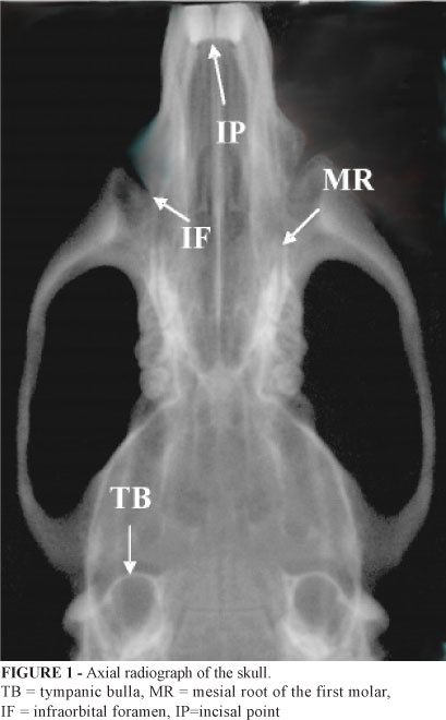
The radiographs were subjected to computerized cephalometric evaluation. The radiographic images were digitized using an optical reader. Measurements were obtained using the Imagelab software. Using axial skull radiographs, the following distances were measured bilaterally: TB-MR - tympanic bulla to the mesial root of the first molar; TB-IF - tympanic bulla to infraorbital foramen; and IF-IP - infraorbital foramen to incisal point (Figure 1). On the lateral radiographs of the hemimandibles, the following distances were measured bilaterally: CP-AP - condylar process to angular process; TM-AN - from the intersection of the distal face of the third molar with the mandibular ramus to the antegonial notch (on the base of the mandible, anteriorly to the mandibular angle); II-CP - lower insertion of incisor to condylar process; and II-AP - lower insertion of incisor to angular process (Figure 2).
To evaluate the significance of differences between the mean values from the right and left sides in each group, the paired Student's t-test was used, while analysis of variance (ANOVA) and Tukey's test were used for the mean values of the three groups. The significance level was set at 5% (p < 0.050).
Results
Macroscopic examination of the specimens revealed facial asymmetry in the experimental group, with deviation of the mandible towards the operated side. The right-side medial pterygoid and masseter muscles were seen to present smaller volumes in the experimental group. Contour alterations with a smaller angular process were noted radiographically only in the experimental group.
The mean values of distances found from the axial radiographs of the skulls are presented in Table 1. From ANOVA, there were no significant differences between any of the measurements.
The mean values for the distances found from the lateral radiographs of the hemimandibles are presented in Table 2. From ANOVA, there were significant differences in II-AP (p < 0.001), and TM-AN (p = 0.008) on the right side. Next, using Tukey's test, it was demonstrated that for II-AP, the mean value in the experimental group was smaller than in the sham-operated group (p = 0.002) and in the control group (p=0.001), while for TM-AN the mean value in the sham-operated group was smaller than in the experimental group (p = 0.028) and in the control group (p = 0.012). Lastly, using the paired Student's t-test to compare the sides of each group, significant differences were only found for II-AP in the experimental group (p < 0.001) and TM-AN in the sham-operated group (p = 0.043), thus confirming the previous findings.
Discussion
The effects of detachment and repositioning of the medial pterygoid muscle on the growth of the maxilla and mandible were analyzed in young rats. Thus, when the animals reached adult age, radiographic projections were obtained and were used to perform cephalometry on a computer system. Through statistical analyses, it could be seen that changes occurred only in the mandible. The regions that presented growth abnormalities caused by the detachment of the medial pterygoid muscle could be identified.
Macroscopic evaluation revealed facial asymmetry with deviation of the median line of the mandible towards the operated side and smaller volume of the medial pterygoid and master muscles in the experimental group. The smaller volume of these muscles indicates that this was a consequence of the surgical procedure. Surgical access at young ages may have negative effects on the growth of the maxilla and mandible4,5.
Detachment of the periosteum and musculature by means of surgical access may result in size or shape alterations to the maxilla and/or mandible5,8. The periosteum influences mandibular growth, and a migration pattern has been observed9. The possibility of decreased mandibular growth caused by the periosteal injury needs to be considered10. However, other studies have suggested that periosteal detachment alone or surgical access does not cause growth abnormalities and, if such abnormalities occur, they are minor in nature and temporary11,12.
Cephalometric evaluations from radiographs on the skull or hemimandibles of dissected specimens using a computer system, as in this study, lead to reliable measurements13. The distances in this study were similar to those in other studies4,14,15,16.
The abnormalities found in this study were located on the mandible, particularly in the posterior regions of the body and angle. Thus, radiographically observed abnormalities were confirmed by cephalometry. The smaller length of the mandible relative to the angular process in the experimental group was the main finding, thereby indicating the action of the medial pterygoid muscle on the growth and shape of the mandibular angle region. This suggests that there was a lack of ability to displace the osseous area to which the muscle adheres, towards the muscle origins and insertions1. However, another important finding was that the height of the mandibular body was smaller in the sham-operated group. A similar abnormality was expected in the experimental group, which indicates the need for new studies.
It has been reported in the literature that the action of the masticatory muscles on mandibular growth in rats is intense in the regions of the angular and condylar processes17. Similar processes are found in monkeys, with bone apposition on the posterior, inferior and lateral borders of the mandible18. Thus, studies have demonstrated that the lateral pterygoid muscle has an effect on the growth of the condylar process19,20; the masseter muscle has an effect on the growth of the angular process2,6; and the temporalis muscle has an effect on the growth of the coronoid process and mandibular ramus2.
No growth abnormalities were observed in the maxilla in this study. This finding is probably due to compensation for muscle activity and the intense adaptive capability consequent to extrinsic mechanical factors, which results in growth compensation21. Other studies inducing condylectomy or fracture of the mandibular body may have promoted abnormalities in the maxilla that could be explained by the influence of occlusal intercuspation1,4,5.
Conclusion
Detachment and repositioning of the medial pterygoid muscle in young rats had consequences on the growth of the angle region of the mandible. There were contour changes with significant shortening of the hemimandibles in the experimental group, while the sham-operated group presented smaller height of the body region.
Received: November 25, 2008
Review: January 12, 2009
Accepted: February 18, 2009
Conflict of interest: none
Financial source: FUNDECTO
How to cite this article
Cruz DZ, Rodrigues L, Luz JGC. Effects of detachment and repositioning of the medial pterygoid muscle on the growth of the maxilla and mandible of young rats. Acta Cir Bras. [serial on the Internet] 2009 Mar-Apr;24(2). Available from URL: http://www.scielo.br/acb
- 1. Oyen O J. A função mastigatória e o crescimento e desenvolvimento facial In: Enlow DH, Crescimento facial. 3ed. São Paulo: Artes Médicas; 1993. p. 272-90.
- 2. Lifshitz J. Comparative anatomic study of mandibular growth in rats after bilateral resection of superficial masseter, posterior temporal, and anterior digastric muscles. J Dent Res. 1976;55:854-8.
- 3. Luz JGC, Araújo VC. Rotated subcondylar process fracture in the growing animal: an experimental study in rats. Int J Oral Maxillofac Surg. 2001;30:545-9.
- 4. Rocha EMVF, Goulart AC, Goldenberg S, Luz JGC. Efeitos da fratura de corpo da mandíbula no crescimento da maxila e da mandíbula: estudo experimental em ratos jovens. Acta Cir Bras. 2002;17:102-9.
- 5. Rodrigues L, Luz JGC. Conseqüências da remoção do côndilo mandibular no crescimento da maxila e da mandíbula: estudo experimental em ratos. Acta Cir Bras. 2001;16:15-25.
- 6. Avis V. The significance of the angle of the mandible: an experimental and comparative study. Am J Phys Anthrop. 1961;19:55-61.
- 7. Guelinckx P, Dechow PC, Vanrusselt R, Carlson DS. Adaptations in the temporalis muscle of rabbits after masseter muscles removal. J Dent Res. 1986;65:1294-9.
- 8. Schweinfurth JM, Koltai PJ. Pediatric mandibular fractures. Facial Plastic Surg. 1998;14:31-44.
- 9. Frankenhuis-Van Den Heuvel THM, Maltha JC, Kuijpers-Jagtman AM, Van't Hof MA. A longitudinal radiographic study of the periosteal migration along the growing rabbit mandible. J Dent Res. 1992;71:398-402.
- 10. Koski K, Rönning O. Condyle neck periostomy and the mitotic activity in the condylar tissues of the young rats. Swed Dent J. 1982;15:109-13.
- 11. Sa'do B, Tashiro H. The influence of periosteal elevation on mandibular growth in young rats. Int J Oral Maxillofac Surg. 1989,18:175-8.
- 12. Cabezas NT. Aspectos do crescimento facial de coelhos, com fraturas mandibulares, após osteossínteses com microplacas de titânio [Tese Doutorado]. São Paulo: Universidade de São Paulo, Faculdade de Odontologia; 1996.
- 13. Del Campo AI, Elizondo MM, Magnelli LM, Valadez AS, Ontiveros DS. Craniofacial development in rats with early resection of the zygomatic arch. Plast Reconstr Surg. 1995;95(3):486-95.
- 14. Yamamoto MK, Novelli MD, Luz JGC. Effects of unilateral upper incisor extraction on facial growth of young rats. J Nihon Univ Dent Sch. 1997;39:191-5.
- 15. Rodrigues L, Miyazaki, L T, Luz J C. Conseqüências no crescimento de maxila e mandíbula de defeito ósseo cirúrgico no ramo da mandíbula de ratos. Acta Cir Bras. 2004;19:523-8.
- 16. Teixeira VCB, Teixeira ACB, Luz JGC. Skeletal changes after experimentally displaced condylar process fracture in growing rats. J Cranio-Maxillofac Surg. 2006;34:220-5.
- 17. Kiliaridis S. Muscle function as a determinant of manfibular growth in normal and hypocalcaemic rat. Eur J Orthod. 1989;11:298-308.
- 18. Turpin, D. L. Growth and remodeling of the mandible in the Macaca mulatta monkey. Am J Orthod. 1968;54:251-71.
- 19. Whetten LL, Johnston LE Jr. The control of condylar growth: An experimental evaluation of the role of the lateral pterygoid muscle. Am J Orthod. 1985;88:181-9.
- 20. Stutzmann J J, Petrovic AG. Role of the lateral pterygoid muscle and menisco temporomandibular frenum in spontaneous growth of the mandibule and in growth stimulated by the postural hyperpropulsor. Am J Orthod Dentofacial Orthop. 1990;97:381-91.
- 21. Koski K. Cranial growth centers: facts or fallacies? Am J Orthod. 1968;54:566-83.
Publication Dates
-
Publication in this collection
08 Apr 2009 -
Date of issue
Apr 2009
History
-
Accepted
18 Feb 2009 -
Reviewed
12 Jan 2009 -
Received
25 Nov 2008