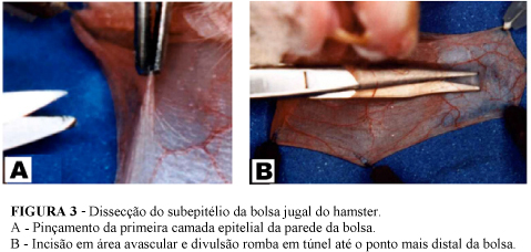
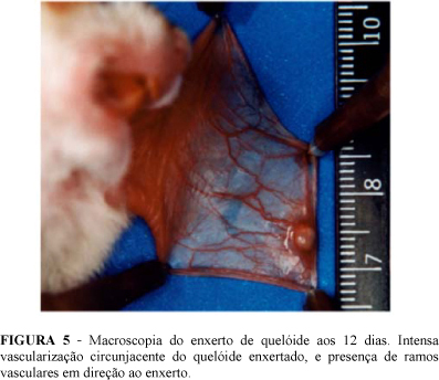
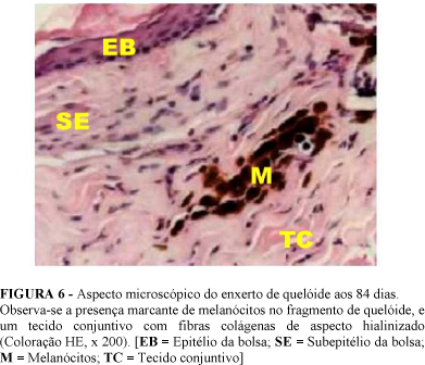
PURPOSE: To describe the integration of keloid heterograft into hamster (Mesocricetus auratus) sub-epithelium. METHODS: The sample is formed by 18 male hamsters, not isogenetic ones, aged between 10 and 14 weeks. Keloid fragments were obtained from keloid scars of the breast region of adult female mulatto patient. Each hamster received keloid fragments into both of its pouches, in a total of 36 grafted fragments. Animals were distributed into 6 groups for having their grafts assessed in the days 5, 12, 21, 42, 84, and 168. A macroscopic assessment is performed by comparing the pouch containing the grafted fragment, at each time point, with the same pouch in the immediate post surgical moment through a comparison of standardized photographs. Under microscope, the presence of blood vases is considered within the conjunctive tissue of the grafted fragment, as a criterion of its integration. Other events, as keratin secretion, the presence of cellular infiltrated, and epithelium and keloid collagen fibers aspects are also analyzed. RESULTS: Macroscopy reveals intensive vascularization of the pouch up to 12 days from the transplant, and the presence of constant dark brown pigmentation on the grafted keloid fragments. In microscopy the integration of keloid fragments is found by the presence of blood capillary vases within conjunctive tissue. The presence of intensive cellular inflammatory type infiltrated up to 12 days is also observed, as well as the remaining of keloid epithelium up to 21 days, and the appearing of melanocytes from the day 42. CONCLUSION: Hamster cheek pouch represents, a priori, an experimental model for the investigation of keloid.
Mesocricetus; Transplantation; Skin transplantation; Keloid


