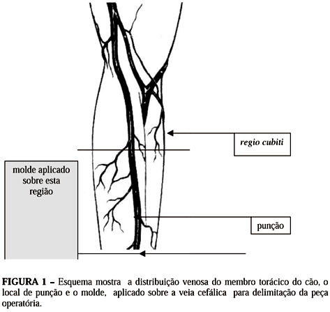
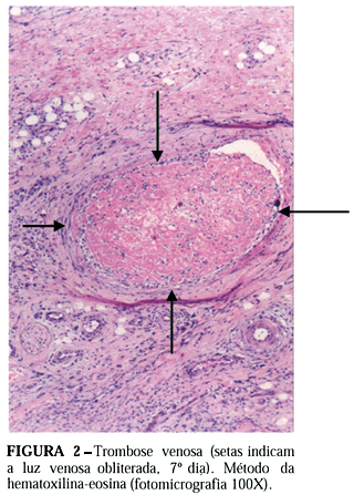
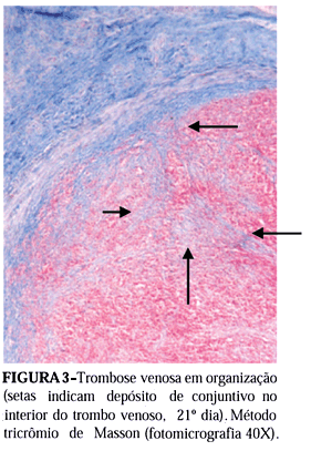
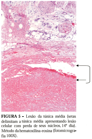
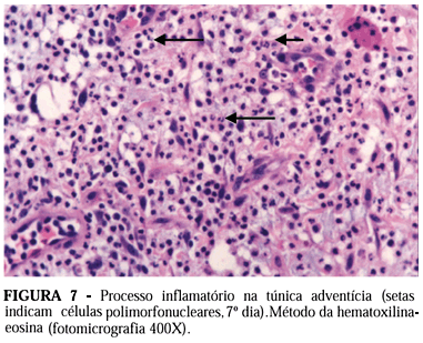
OBJECTIVE: Evaluate the ethanolamine oleate effects on venous dog wall. METHODS: The cephalic vein wall changes were evaluated in 39 male adults mongrel dogs, weighing 10-18 kg, randomly distributed in three groups (group 1 = 7 days, group 2 = 14 days and group 3, 21 days). Single punction and injection of 2 mL of 5 % ethanolamine oleate and 7, 14 and 21 days later operative specimen excision were compared to non-injected contralateral control vein. Histological parameters evaluated (hematoxylin-eosin and Masson's trichrome staining methods) were: venous thrombosis and organization, thrombus recanalization, media layer lesion and inflammatory process, outer wall inflammatory process, hemossiderin, sclerosant spillage outside the outer layer and hyaline amorphous material deposition. RESULTS: Venous thrombosis and thrombus organization were seen in all animals. Thrombus recanalization was not shown until 21 days. Media layer lesion occurred without inflammatory process. Outer wall inflammatory process was seen in all three time periods. Hemossiderin phagocytes occurred on 14th and 21st days. Sclerosant spillage outside the outer layer was seen only on the 7th day. Hyaline amorphous material deposition was seen only on the 21st day. CONCLUSIONS: Ethanolamin oleate in contact with the inner vein wall produced venous thrombosis, organized in all cases. During this study no significant recanalization was observed. Media layer vein lesion was seen in all animals without any correlate inflammatory reactive process. Reactive inflammatory process, hemossiderin phagocytosis, sclerosant spillage and hyaline amorphous material deposition was shown in the adventitia layer.
Veins; Ethanolamine oleate; Dogs; Sclerosing solutions; Sclerotherapy

