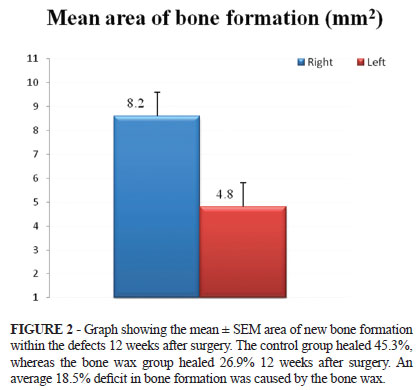
PURPOSE: To quantify the amount of bone formation in the calvarial region of Wistar rats after craniotomy using bone wax as a haemostatic agent. METHODS: Surgery to produce bilateral, symmetric, full-thickness cranial defects (area: 18 mm²) was performed in eight animals. The right side of the cranium remained open and the edges of the left side osseous defect was covered with bone wax. Calvaria were imaged immediately after surgery and 12 weeks postoperatively by computerized tomography. The areas of the bone defects were measured in three-dimensional images using Magics 13.0 (Materialise-Belgic, software CAD). RESULTS: The average amount of bone formation on the left and right side respectively was 4.85 mm² and 8.16 mm². Statistically significant differences between the amount of bone formation on the left and right sides were seen. CONCLUSIONS: Bone wax significantly diminishes the rate of bone formation in calvarial defects in a rat model.
Waxes; Skull; Hemostatics; Osteogenesis; Rats