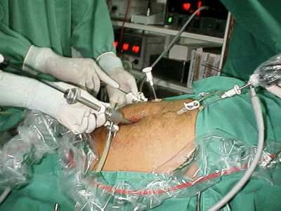
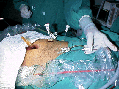
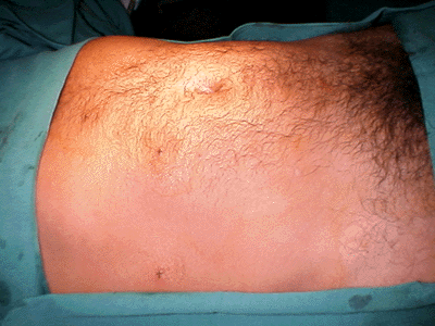
The authors describe the technique they employed to perform needlescopic cholecystectomies in the Núcleo de Hospital de Aeronáutica de Manaus, from September 1997 to May 1999. A complete videolaparoscopic set of equipment was used in the procedure in association to an extra videocapture set for the microlaparoscope (video monitor, microcamera and light source). The procedure is mainly performed with the needlescopic instruments (MiniSite®) inserted in the epigastric, upper right abdominal quadrant and right flanc 2mm trocar sleeves, introduced through the anterior abdominal wall with 10 mm umbilical videolaparoscopic guidance. The microlaparoscope was employed through the epigastric port to monitor the 10 mm clip applier, inserted through the umbilical port to ligate the cistic elements. Likewise, it was employed to monitor gallbladder removal from the abdominal cavity through the umbilical wound. The 2 mm skin wounds were closed with sterile surgical tape. A total of 83 patients (11 males, 72 females), ranging from 17 to 87 years of age, with gallbladder diseases were operated. There was no patient selection in the study but most of the cases (89.2%) did not present inspissated gallbladder walls. The mean operative time was 92 ± 21 min and the average time of hospitalization was 16 h. The main peroperative incident observed was gallbladder puncture with bile leakage to the abdominal cavity (41%), which was considered due to the surgical team’s learning curve in the method. Vomits were the main postoperative complication (51.8%) and there was no instance of wound infection. The complete method of needlescopic cholecystectomy was employed in 82% of the patients; in 6% of the patients, a 5 or 10 mm portal had to be put in place of one of the 2 mm to deal with thick walled gallbladders or to correct image problems encountered with the 2 mm laparoscope; in another 6% of the patients, an extra 10 mm suprapubic portal was employed to receive the 10 mm laparoscope when the umbilical portal was used with 5 and 10 mm instruments (in these cases the 2 mm laparoscope was not available due to fiberoptic deterioration). In 3.6 % of the surgeries, the needlescopic method had to be converted to the usual videolaparoscopic one (with two 5 mm and two 10 mm portals). Two operations had to be converted to conventional cholecystectomies (one due to equipment failure and the other due to a solid block of inflammatory adhesions to the hepatic hilar structures). The authors conclude that needlescopic cholecystectomies are feasible, demand the surgical team to go through a new learning curve period and are responsible for a longer operative time due to peculiarities of the instruments and equipment involved.
Cholecystectomy; Laparoscopy; Microlaparoscopy; Minimally invasive surgery


