Klebsiella pneumoniae isolated from the intestines of Tenebrio molitor larvae (Coleoptera: Tenebrionidae) that consume expanded polystyrene
Abstract
Plastics such as polystyrene are resistant to biodegradation, pollute the environment, and negatively impact the health of living organisms. However, several organisms, such as the larvae of Tenebrio molitor (Coleoptera: Tenebrionidae) and their associated gut microbiome, contribute to its degradation. The aim of this research was to determine the efficiency of degradation of expanded polystyrene (EPS) by gut bacteria isolated from larvae of T. molitor. To achieve this, a set of EPS-degrading bacteria was selected based on the time required to utilize the polymer as a carbon and energy source. Additionally, EPS degradation efficiency was compared, and the most efficient degrading bacterium was identified at the molecular level. Results showed that 95.13% of the bacteria isolated on nutrient agar and 86.57% of those isolated on MacConkey agar were able to grow on EPS. Five selected bacteria were able to degrade the polymer after 36 hours of incubation. The efficiency of EPS degradation, expressed as the percentage of weight loss by the degrading bacteria, ranged from 5.29% to 12.68%, with a reduction rate of 0.0005 to 0.0013 g per day and a half-life of 533.15 to 1386.20 days. Finally, 16S rRNA gene analysis identified the bacterium as Klebsiella pneumoniae. Cultivable gut bacteria from T. molitor larvae have demonstrated potential as candidates for EPS degradation, and biotechnological techniques can further enhance the efficiency of the degradation process.
Keywords:
biotechnological techniques; gut microbiome; polystyrene degradation
Resumo
Plásticos como o poliestireno são resistentes à biodegradação, poluem o ambiente e impactam a saúde dos seres vivos; no entanto, vários organismos, como as larvas de Tenebrio molitor (Coleoptera: Tenebrionidae) e seu microbioma intestinal associado, contribuem para sua degradação. O objetivo desta investigação foi determinar a eficiência da degradação do poliestireno expandido (EPS) por bactérias intestinais isoladas de larvas de T. molitor. Para isso, foi selecionado um conjunto de bactérias degradadoras de EPS de acordo com o tempo necessário para a utilização do polímero como fonte de carbono e energia. Além disso, a eficiência da degradação do EPS foi comparada, e as bactérias com maior eficiência de degradação foram identificadas a nível molecular. Verificou-se que 95,13% das bactérias isoladas em ágar nutriente e 86,57% das isoladas em ágar MacConkey cresceram com EPS, selecionando-se cinco bactérias que utilizaram o polímero após 36 horas de incubação. A eficiência da degradação do EPS, expressa em porcentagem de peso perdido pelas bactérias degradadoras, foi de 5,29% a 12,68%, com uma taxa de redução de 0,0005 a 0,0013 g/dia e uma meia-vida de 533,15 a 1386,20 dias. Finalmente, o gene do 16S rRNA identificou a bactéria como Klebsiella pneumoniae. As bactérias cultivadas do intestino de larvas de T. molitor provaram ser potenciais candidatas à degradação do EPS, e técnicas biotecnológicas aumentarão a eficiência desse processo.
Palavras-chave:
degradação de poliestireno; microbioma intestinal; técnicas biotecnológicas
1. INTRODUCTION
Petroleum-derived plastics, or petroplastics, are components of most everyday materials due to their lightweight, durability, and low cost. As a result, global production has increased exponentially, reaching over 390.7 million metric tons (MT) per year, compared to 1.5 million MT produced in 1950 (Shukla et al., 2024; USAID, 2020). Most plastics are single-use and do not degrade easily, leading to an estimated 4.9 billion MT discarded between 1950 and 2005 that persist somewhere in the world (Song et al., 2020). Petroplastics in the form of macroplastics (>25 mm), mesoplastics (5-25 mm), large microplastics (1-5 mm), small microplastics (20 µm-1 mm), and nanoplastics (1-1000 nm) contaminate soil, water, and air, impacting the health of all living beings (Shukla et al., 2024; Hwang et al., 2020).
Polystyrene (PS) is a polymer with an amorphous structure, high rigidity, and lightweight properties, making it low-cost and fully recyclable. However, the recycling market is not significant. Additionally, it is marketed in various forms, including expanded polystyrene (EPS) (Song et al., 2020). Due to poor segregation of EPS, it is found contaminating various levels of the ecosystem. Oceans contain high amounts of EPS, which, when exposed to sunlight, fragments and generates microplastics and nanoplastics (Du et al., 2024). PS has also been detected in soil at depths of 0-30 cm in countries such as Germany, China, and Spain (Dioses et al., 2020). Furthermore, PS has been reported as an air pollutant in studies conducted in Paris, London, and Dongguan (Amato et al., 2020). In Peru, pollution from plastics such as PS (De la Torre et al., 2024) is increasing daily, with more than 500 microplastics per square meter recorded on the beaches of Lima and Callao (USAID, 2020).
In living organisms, the presence of PS in the soil has been shown to alter photosynthesis (Zhuang et al., 2024), reduce chlorophyll (Iswahyudi et al., 2024), carotenoids, micronutrients, as well as plant growth and biomass (Lian et al., 2021). In animals, PS can reduce sperm quality and serum reproductive hormones (Ying et al., 2024), alter ovarian function, decreasing oocyte quality and fertility (Xue et al., 2024), and cause dysregulation of genes associated with neurotransmitters in embryos (Shubham et al., 2024). Additionally, it can increase enzymes related to oxidative stress (Zhen et al., 2024), cause toxicity in hemolymph and digestive glands (Ventura et al., 2024), and increase alcohol/aldehyde dehydrogenase (AADH) and lactate dehydrogenase (LDH) enzymes in tissues, reducing survival (Gagné et al., 2023). On the other hand, it can reduce the rate of photosynthesis and nitrogen fixation in cyanobacteria (Lixia et al., 2024).
Humans ingest styrene monomers from various sources such as food, water, medical devices, and everyday products. It has been estimated that human consumption of PS can exceed 133 mg per year when particles have a diameter of 100 µm (Hwang et al., 2020). PS nanoparticles (25-70 nm in diameter) can be absorbed by living organisms, significantly affecting the viability of human alveolar epithelial cells, activating genes related to the inflammatory response, modifying the expression of proteins associated with the cell cycle, and inducing pro-apoptosis (Xu et al., 2019).
In nature, various larval forms of insects, such as Tenebrio molitor Linnaeus, 1758 (Yang et al., 2023), T. obscurus (Peng et al., 2019), Uloma sp. (Kundungal et al., 2021), Tribolium castaneum (Rodríguez et al., 2021), Zophobas atratus (Lu et al., 2024), Z. morio (Sun et al., 2022), Galleria mellonella (Young et al., 2024), and Alphitobius diaperinus (Cucini et al., 2022), are known to fragment polystyrene (PS) to consume and ingest it into their intestines. In the intestines, PS is ultimately mineralized by specialized intestinal bacteria, including Acinetobacter, Alcaligenes, Bacillus, Citrobacter, Enterobacter, Klebsiella, Massilia, Pseudomonas, and Serratia (Sufang et al., 2024; Machona et al., 2022; Vera et al., 2021; Tan et al., 2021a; Urbanek et al., 2020; Jiang et al., 2021; Brandon et al., 2021). These bacteria degrade the polymer into carbon dioxide and water, presenting a potential alternative for reducing this pollutant (Wang et al., 2020; Peng et al., 2019). Therefore, the objective of this study was to determine the efficiency of expanded polystyrene degradation by cultivable bacteria associated with the intestines of Tenebrio molitor larvae (Coleoptera: Tenebrionidae).
2. MATERIAL AND METHODS
2.1. Preparation of emulsified EPS for feeding T. molitor larvae
The T. molitor larvae, with an average length of 17-18 mm, were purchased from the online store Kuru Wasi in Lima, Peru, while the EPS was obtained from ETSAPOL, SKU: 11760. To prepare the emulsified EPS, 3 g of EPS were placed in a beaker and dissolved in 97 mL of dichloromethane (Spectrum), then allowed to rest at 4°C for 48 hours. The emulsified EPS sheets adhered to Petri dishes were carefully removed, cut into 1 cm² fragments, and placed in tubes with pre-sterilized carbon-free minimal basal broth (Tan et al., 2021b). The wheat bran was sterilized at 120°C for 10 minutes and then dried at 80°C for 10 minutes, while the EPS was sterilized at 80°C for 10 minutes. Both were cooled to room temperature before being fed to the larvae (Bae et al., 2021).
2.2. Quantification of cultivable bacteria associated with the intestinal tract of T. molitor larvae
The number of colony-forming units (CFU mL⁻¹) of bacteria from the intestinal content was determined in 60 larvae of T. molitor. Three different feeding conditions were used for each group of 20 larvae: A) Directly from the commercialization center (LCC), which were fed with slices of potatoes, carrots, and wheat bran (15.96 g); B) From a laboratory rearing with a mixed diet of potatoes, carrots, wheat bran and EPS (1:1:2:2) (LRMD); and C) From a rearing with a diet consisting solely of EPS (1.61 g) (LREPS) over a period of 21 days (Tan et al., 2021a). The rearing temperature, irrespective of the food source, ranged from 21 to 29 °C, with a relative humidity of 53% to 59%. The larvae, with an average length of 20-24 mm, (20 from LCC, 20 from LRMD, and 20 from LREPS) were first chilled at -4°C for 15 minutes to inactivate them (Vera et al., 2021). They were then disinfected by immersion in 75% ethanol for 1 minute, followed by rinsing twice with 0.85% NaCl solution (p/v) (Tan et al., 2021a) and dried with sterile filter paper. Each larva was placed on a Petri dish, and pressure was applied to the posterior region to obtain the intestines, which was then placed in a tube with 1 mL of 0.85% NaCl solution (p/v). Decimal dilutions up to 10⁻⁴ were prepared from this tissue, and these dilutions were plated in three separate replicates on Plate Count agar to enumerate the CFU after incubation in aerobic conditions at 30°C for 24 hours (Urbanek et al., 2020).
2.3. Selection of EPS-emulsifying degrading bacteria
Isolation of bacteria was performed from both the 10⁻¹ diluted and undiluted intestinal content of each T. molitor larva. For the enrichment of the 10⁻¹ dilution, a 0.5 mL aliquot was transferred to tubes containing 4.5 mL of carbon-free minimal basal broth and a 1 cm² fragment of emulsified EPS. The tubes were incubated at 30°C for 7 days and at 25°C for 21 days (Tan et al., 2021b). Subsequently, aliquots from both the enriched and unenriched dilutions were plated by dilution and streaking on nutrient agar and MacConkey agar. After incubation at 30°C for 48 hours, representative bacterial colony morphotypes were selected and subcultured on Tryptic Soy Agar (TSA) in duplicate, followed by Gram staining. The identification of bacterial genera was performed using a biochemical test panel that included triple sugar iron agar (TSI), lysine iron agar (LIA), citrate agar, urea agar, methyl red-Voges Proskauer broth, and tryptophan broth. Pure cultures were stored at 4°C.
To select EPS-degrading bacteria, they were cultured in duplicate in carbon-free minimal basal broth with a 1 cm² fragment of emulsified EPS as the carbon source, and 1% glucose as a positive control. After incubation at 30°C for 7 days, the optical density of the cultures was measured using a visible light spectrophotometer (Model Tenso Med NV-203) at 600 nm, with non-inoculated EPS and glucose broth as the blank. The ten bacteria isolated on nutrient agar and the ten isolated on MacConkey agar that exhibited the highest optical density values in the minimal basal broth with EPS were selected. These were then plated in 5 mL of the same broth with EPS and 20 µL of a 1% solution of 2,3,5-triphenyltetrazolium chloride (TTC) as a viability indicator. The cultures were incubated at 30°C, with readings taken every 12 hours to observe any color change in the indicator to red, which would indicate metabolic activity and thus the utilization of EPS as a carbon and energy source (Tan et al., 2021b).
2.4. Comparison of the efficiency in the degradation of emulsified EPS
The efficiency of EPS degradation by the selected bacteria was indirectly determined by measuring the turbidity in the culture medium, which correlates with microbial growth; and directly by A) assessing the weight loss of the polymer or substrate consumed (Maroof et al., 2021), B) the reduction rate (k), and C) the half-life of the residual EPS (t₁/₂) (Auta et al., 2017). To determine turbidity, 3% of a 12-hour bacterial inoculum was added to 50 mL of minimal basal broth, along with three 1 cm² fragments of emulsified EPS. The mixture was incubated at 30°C, and its optical density at 600 nm was measured every 12 hours for 72 hours, comparing it with an uninoculated control and a control with glucose (Tang et al., 2017).
The weight loss of the polymer was determined by culturing the selected bacteria in triplicate flasks containing 100 mL of minimal basal medium and 0.36 - 0.48 g of emulsified EPS (initial weight). After incubation at 30°C for 100 days, the polymer was extracted and mixed with sodium dodecyl sulfate, then incubated at 50°C with constant agitation (40 rpm) to remove adhered bacterial cells (Wang et al., 2020). The polymer was then washed with distilled water and dehydrated at 60°C until a constant weight (final weight) was achieved. The percentage of weight loss was calculated based on this (Jiang et al., 2021).
The reduction rate (k) was calculated using the weight loss (Auta et al., 2017) (Equation 1):
Where W = Final weight y W0 = Initial weight
The half-life of the residual EPS (t₁/₂) (Auta et al., 2017) was calculated as follows (Equation 2):
2.5. Molecular characterization of the microbial strain
Bacterial DNA extraction was carried out using the Wizard Genomic DNA Purification Kit (Promega). The 16S rRNA gene was amplified by PCR with the universal primers 27F and 1492R. The PCR products were then sequenced using the Sanger method at the “Marcel Gutiérrez Correa” Mycology and Biotechnology Laboratory at the Universidad Nacional Agraria La Molina (UNALM). The assembled sequences were preliminarily identified using BLASTN and aligned with related 16S rRNA gene sequences retrieved from the NCBI database (Altschul et al., 1997). Finally, phylogenetic analysis was performed using MEGA11 (Tamura et al., 2021), employing the Neighbor-Joining method and 1000 bootstrap replicates.
2.6. Data processing and statistical analysis
Bacterial efficiency in the degradation of PS, expressed as the reduction rate and half-life of residual PS, is presented as mean values with standard deviation (±). The mean weight loss due to the use of EPS as a carbon and energy source was analyzed using parametric statistics to determine differences between treatments (analysis of variance) and their significance (Tukey's multiple comparison test), with a significance level of 0.05. IBM SPSS Statistics Version 28 was used for the analysis.
3. RESULTS AND DISCUSSION
3.1. Number of cultivable bacteria associated with the intestines of Tenebrio molitor larvae
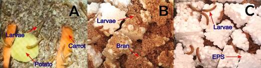
The average cultivable bacteria associated with the intestines of 20 larvae varied depending on the diet provided (Table 1, Figure 1).
Average cultivable bacteria (CFU mL-1) associated associated with the intestines of Tenebrio molitor larvae.

T. molitor larvae from the commercialization center (A), from laboratory rearing with a mixed diet of wheat bran and EPS (B), and from rearing with a diet consisting solely of EPS (C).
The hydrophobicity of most plastics, such as EPS, makes them resistant to hydrolysis, which negatively impacts their chemical or biological degradation (Ho et al., 2017). Despite this, T. molitor larvae from the Tenebrionidae family were able to consume EPS for 21 days, consistent with reports by Cunguan et al. (2023), Vera et al. (2021), and Brandon et al. (2021). EPS consumption was confirmed by the weight loss of the polymer, which indicates its use as a carbon source (Machona et al., 2022).
Microorganisms associated with the larvae's intestines play a crucial role in the catabolism of EPS (Tsochatzis et al., 2021), without diminishing the larvae's own activity in fragmenting the polymer into smaller particles (Machona et al., 2022). Brandon et al. (2021) reported no significant difference in the activity of the supernatant from the intestinal content of T. molitor larvae fed wheat bran or polystyrene (PS), both supplemented with antibiotics. In both cases, emulsification activity was observed, suggesting that fragmentation of the polymer into smaller particles is inherent to the larvae's intestines, regardless of the type of food consumed. Jiang et al. (2021) concluded that larvae and intestinal microorganisms have a symbiotic interaction. After ingesting plastic, intestinal microorganisms play a role in biodegradation, but the larvae’s enzymatic system also contributes to the degradation process through chewing, which increases the specific surface area and promotes contact with the extracellular enzymes in their intestinal tract. Moreover, Przemieniecki et al. (2020) investigated changes in the enzymatic profile of digestive tract lysates from T. molitor larvae and PS-degrading intestinal microorganisms, identifying enhanced enzymatic activity associated with enzymes such as alkaline and acid phosphatases, esterase, lipase, galactosidase, glucuronidase, glucosidase, and mannosidase.
The number of bacteria was lower in the intestines of T. molitor larvae fed EPS compared to larvae from the commercialization center and those fed a mixed diet. This result is consistent with the findings of Tsochatzis et al. (2021), who demonstrated that the number of bacteria in the intestines of T. molitor larvae fed a wheat bran: PS diet at a 4:1 ratio was lower than that of larvae fed a wheat bran diet (control). This difference was attributed to the bacteriostatic effect of PS degradation products, including styrene oligomers. Additionally, Urbanek et al. (2020) showed that the highest number of colony-forming units of bacteria was found in the intestines of the control group larvae. This number varied depending on the type of PS, being higher with PS used for packaging (44.325 × 10⁶) compared to EPS, which has a molecular weight ratio (MFR) of polymer chain length 3.5 times greater.
3.2. Selected EPS-emulsifying bacteria
The range of colony morphotypes developed on nutrient agar was from one to four, resulting in 131 pure cultures from the non-enriched culture and 136 from the enriched culture. On MacConkey agar, the range of colony morphotypes was from one to five, yielding 102 pure cultures from the non-enriched culture and 114 from the enriched culture.
Among the 483 cultivable bacteria associated with the intestines of T. molitor larvae, 61.28% (296) were Gram-negative, while 38.72% (187) were Gram-positive. In basal minimal medium free of carbon with a fragment (1 cm²) of emulsified EPS, 95.13% (254) of the bacteria isolated on nutrient agar grew, and 86.57% (187) of the bacteria isolated on MacConkey agar grew. In total, 91.30% (441) of the bacteria grew with EPS, including 59.64% (263) Gram-negative and 40.36% (178) Gram-positive bacteria. From these, the 20 strains (ten from each agar type) with the highest optical density values were selected (Table 2).
The isolation of bacteria from the intestines of T. molitor larvae is consistent with the findings of Machona et al. (2022). The enrichment technique was employed because it favors the selection of microorganisms that are more efficient in degrading PS (Brandon et al., 2021). The bacteria associated with the intestines of T. molitor larvae were predominantly Gram-negative, aligning with reports from Machona et al. (2022), Bae et al. (2021), and Peña et al. (2020). Additionally, it has been observed that the intestinal microbiome of T. molitor larvae is predominantly composed of the phyla Proteobacteria and Firmicutes (Urbanek et al., 2020).
Bacteria that grew using EPS as a carbon source (91.30%) were classified as polymer degraders. Plastic biodegradation begins with the growth of microorganisms on the polymer surface, where they secrete enzymes to break it down into small fragments called oligomers and, eventually, into monomeric units. The pathways for the catabolism of styrene or EPS components are diverse; however, the most common pathway involves the oxidation of styrene to phenylacetate via the citric acid cycle. Degradation products of EPS include styrene oxide, phenylacetaldehyde, phenylacetic acid, and phenylacetyl coenzyme A. The enzymes involved in this process are styrene monooxygenase (SMO), styrene oxide isomerase (SOI), phenylacetaldehyde dehydrogenase (PAALDH), and phenylacetyl coenzyme A ligase (PACoA) (Ho et al., 2017).
It is believed that PS-degrading bacteria can secrete extracellular oxidative enzymes that fragment PS chains and generate intermediates with C=O bonds. Identifying the key enzymes involved in the depolymerization and degradation of PS is crucial (Mamtimin et al., 2023); however, evidence suggests that EPS-degrading bacteria include Gram-negative members of the Enterobacteriaceae family and Gram-positive genera such as Enterococcus and Bacillus, among others (Sun et al., 2022; Tsochatzis et al., 2021; Tan et al., 2021a; 2021b). The bacteria are capable of achieving high rates of PS degradation in environmental settings lacking other carbon sources. However, if other carbon sources are available alongside PS, the rate of contaminant degradation decreases. This limitation underscores the need for further research into the enzymes involved in the PS degradation process and their potential for its commercial application (Ho et al., 2017).
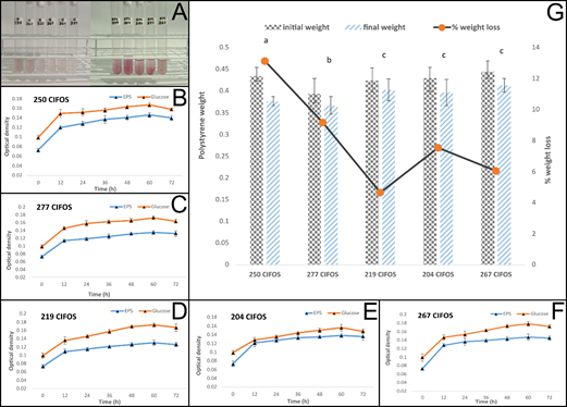
A reduction in the TTC indicator was observed in 65% (13) of the broths cultured with the bacteria (7 from nutrient agar and 6 from MacConkey agar), as evidenced by the appearance of a stable raspberry color after 36-228 hours of incubation (Figure 2A). In the glucose control, the indicator reduction was observed between 24 and 72 hours. Among the TTC-reducing bacteria, the five strains that required 36 hours for TTC reduction while utilizing emulsified EPS as a carbon and energy source were selected.

TTC Indicator (A), optical density of minimal basal broth with glucose and EPS (B, C, D, E, F), and percentage of weight loss of EPS (G) utilized by intestinal bacteria from Tenebrio molitor.
Tan et al. (2021a) demonstrated the reduction of TTC by EPS-degrading bacteria isolated from the intestines of Zophobas morio larvae. The researchers observed TTC reduction from 24 to 96 hours of incubation, considered a primary screening for viability and metabolic activity. This rapid assay verifies EPS degradation when used as a carbon source by the bacteria. TTC, which is initially colorless, is reduced to the colored compound triphenyl formazan (TPF) in the electron transport system. TTC is an artificial electron acceptor and is reduced in the aerobic cytochrome system to form the insoluble and colored compound called formazan (Tan et al., 2021b). The time required for TTC reduction by the intestinal bacteria of T. molitor larvae in the EPS broth (36-228 hours) was longer than in the broths with glucose as the carbon source (24-72 hours). This time difference was also observed by Tan et al. (2021b) with a B. megaterium strain able to degrade PS, concluding that the polymer requires more time to be broken down into short monomers that can enter the bacterial cell and be used as a carbon source during growth.
3.3. Comparison of bacterial efficiency in the degradation of emulsified EPS
The optical density (600 nm) of the broths cultured with bacteria degrading emulsified EPS as a carbon and energy source gradually increased, reaching its maximum density at 60 hours. The timing was similar for the group control with glucose. At 60 hours, the optical density values ranged from 0.130 to 0.147 with EPS and from 0.156 to 0.178 with glucose. The highest optical density values were observed for bacterium 267 CIFOS, which utilized both EPS and glucose as carbon and energy sources (Figure 2B-G). Similarly, Lin and Liu (2021) evaluated the growth of intestinal bacteria from T. molitor and Z. morio larvae, using broth turbidity as an indicator of growth and PS degradation over 48 hours, and quantified CFUs to assess microbial viability.
The efficiency of EPS degradation, expressed as the percentage of weight lost by the degrading bacteria, ranged from 12.68% with bacterium 250 CIFOS to 5.29% with bacterium 267 CIFOS. Reported values in the literature vary, ranging from 7.40% to 12.97% (Jiang et al., 2021; Yang et al., 2015). EPS degradation by microorganisms is evaluated through microscopic observation of changes in its surface structure, such as cracks, fissures, holes, and biofilm formation, as well as alterations in its mechanical and chemical properties (Cucini et al., 2022; Brandon et al., 2021; Tan et al., 2021a). Weight loss of the polymer (gravimetry) provides a direct measure of biodegradation; however, issues such as improper cleaning of the sample or weight loss due to volatilization of intermediate components and soluble impurities may affect the results (Ho et al., 2017).
The polymer degradation by strain 250 CIFOS resulted in a weight loss of 12.68%, which is higher than the 7.46% reported by Auta et al. (2017) for Bacillus cereus. However, the reduction rate in this study was 0.0013 g/day with a half-life of 533.15 days (Table 3), compared to 0.0019 g/day and a residual polymer half-life of 363.16 days reported by Auta et al. (2017). This difference may be explained by the fact that Auta et al. (2017) used UV-irradiated PS for 25 days, a process that increases carbonyl groups and reduces tensile strength, thereby accelerating the rate of reduction and microbial degradation (Crystal et al., 2024).
3.4. Phylogenetic analysis
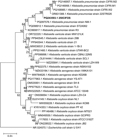
The BLASTN analysis identified the bacterium 250 CIFOS as belonging to the genus Klebsiella, while the phylogenetic analysis confirmed it as K. pneumoniae (GenBank accession number: PQ243593.1) (Figure 3).
It has been demonstrated that bacteria of the genus Klebsiella (family Enterobacteriaceae) remained viable for 28 days in a broth with PS as the carbon source, with their adhesion and growth on the polymer confirmed by scanning electron microscopy (SEM) (Cucini et al., 2022). Additionally, the adhesion of K. grimontii to PS films, as verified by SEM, revealed a reduction in the size and sharpness of the film edges, as well as a deteriorated surface (Park et al., 2023). Furthermore, K. oxytoca, isolated from the intestines of T. molitor larvae that consumed PS, has been reported as a potential polymer degrader by Machona et al. (2022) and Urbanek et al. (2020), similar to K. aerogenes isolated from the intestines of A. diaperinus larvae (Cucini et al., 2022) and from the feces of Z. morio larvae (Sun et al., 2022), and K. variicola isolated from Spodoptera frugiperda larvae (Zhang et al., 2022). In this regard, Sun et al. (2022) concluded that emerging human pathogenic species such as K. oxytoca, Enterococcus spp., and Corynebacterium spp., identified in larvae that were starved and fed with PS, indicated that a diet with EPS is poor and induces an imbalance in the intestinal microbiome; however, these bacteria were associated with PS degradation. On the other hand, K. pneumoniae is an antibiotic-resistant enterobacteria (Araya et al., 2022), an opportunistic pathogen, and a causative agent of both nosocomial and community-acquired infections, especially in immunocompromised patients (Hartantyo et al., 2020). However, this soil-isolated bacterium has been reported as a degrader of microplastics (Saygin and Baysal, 2021) and polyethylene (Zhang et al., 2023). It was also recovered from the intestines of T. molitor larvae fed a diet of bran and PS (in a 4:1 ratio), along with the quantification (µg mg⁻¹) of monomers and oligomers from PS degradation, such as acetophenone (0.180); 2,4-di-tert-butylphenol (0.100); 2,4,6-triphenyl-1-hexane (0.173); 1,1-diphenylethylene (0.016); and styrene (0.005) in the larvae feces (Tsochatzis et al., 2021).
Phylogenetic tree of the 16S rRNA gene for strain 250 CIFOS and other Klebsiella sp. sequences, inferred using the Neighbor-Joining method with 1000 bootstrap replicates. The 16S rRNA sequence of Escherichia coli was used as the outgroup.
4. CONCLUSIONS
Plastic-consuming larvae harbor several microorganisms with potential biotechnological applications. Klebsiella pneumoniae 250 CIFOS demonstrated the highest efficiency in degrading emulsified EPS, with a weight loss of 12.68%, a reduction rate of 0.0013 g/day, and a half-life of 533.15 days. While this bacterium is an opportunistic pathogen, it serves as a valuable source of genes associated with enzymes that promote EPS degradation. Biotechnology offers the potential to accelerate and improve the efficiency of this biodegradation process.
5. REFERENCES
-
ALTSCHUL, S.; MADDEN, T.; SCHAFFER, A.; ZHANG, J.; ZHANG, Z.; MILLER, W. et al Gapped BLAST and PSI- BLAST: a new generation of protein database search programs. Nucleic Acids Research, v. 2, n. 17, p. 3389-3402, 1997. https://doi.org/10.1093/nar/25.17.3389
» https://doi.org/10.1093/nar/25.17.3389 -
AMATO, L.; DOS SANTOS, L.; DE WEGER, L.; HIEMSTRA, P.; VIJVER, M.; MAUAD, T. An emerging class of air pollutants: potential effects of microplastics to respiratory human health? The Science of the Total Environment, v. 749, n. 141676, 2020. https://doi.org/10.1016/j.scitotenv.2020.141676
» https://doi.org/10.1016/j.scitotenv.2020.141676 -
ARAYA, I.; ROACH, F.; TAPIA, T.; RODAS, P.; VILLAMIL, A.; AGÜERO, R. et al Caracterización fenotípica y molecular de cepas de Klebsiella pneumoniae productoras de carbapenemasas tipo OXA-48 circulantes en Chile. Revista chilena de infectología, v. 39, n. 5, p. 551-558, 2022. http://dx.doi.org/10.4067/S0716-10182022000500551
» http://dx.doi.org/10.4067/S0716-10182022000500551 -
AUTA, H.; EMENIKE, C.; FAUZIAH, S. Screening of Bacillus strains isolated from mangrove ecosystems in Peninsular Malaysia for microplastic degradation. Environmental Pollution, v. 231, n. 2, p. 1-8, 2017. https://doi.org/10.1016/j.envpol.2017.09.043
» https://doi.org/10.1016/j.envpol.2017.09.043 -
BAE, J.; CHO, H.; JUNG, H.; PARK, J.; YUN, S.; HA, S. et al Changes in intestinal microbiota due to the expanded polystyrene diet of mealworms (Tenebrio molitor). Indian Journal of Microbiology, v. 61, n. 2, p. 130-136, 2021. https://doi.org/10.1007/s12088-021-00922-w
» https://doi.org/10.1007/s12088-021-00922-w -
BRANDON, A.; GARCIA, A.; KHLYSTOV, N.; WU, W.; CRIDDLE, C. Enhanced bioavailability and microbial biodegradation of polystyrene in an enrichment derived from the gut microbiome of Tenebrio molitor (mealworm larvae). Environmental Science & Technology, v. 55, n. 3, p. 2027-2036, 2021. https://doi.org/10.1021/acs.est.0c04952
» https://doi.org/10.1021/acs.est.0c04952 -
CRYSTAL, X.; LO, S.; RAMANAN, R.; TEY, B.; HUY, N.; WEI, O. Enhancing plastic biodegradation process: strategies and opportunities. Critical Reviews in Biotechnology, v. 44, n. 3, p. 477-494. 2024. https://doi.org/10.1080/07388551.2023.2170861
» https://doi.org/10.1080/07388551.2023.2170861 -
CUCINI, C.; FUNARI, R.; MERCATI, D.; NARDI, F.; CARAPELLI, A.; MARRI, L. Polystyrene shaping effect on the enriched bacterial community from the plastic-eating Alphitobius diaperinus (Insecta: Coleoptera). Symbiosis, v. 36, p. 305-313, 2022. https://doi.org/10.1007/s13199-022-00847-y
» https://doi.org/10.1007/s13199-022-00847-y -
CUNGUAN, J.; ROJAS, L.; MOROCHO, T.; ARCOS, B.; ORTIZ, C. Biodegradation of plastic with Tenebrio molitor insect larvae as an interdisciplinary contribution to environmental biotechnology. Revista Ciencia UNEMI, v. 16, n. 41, p. 28-33, 2023. https://doi.org/10.29076/issn.2528-7737vol16iss41.2023pp28-33p
» https://doi.org/10.29076/issn.2528-7737vol16iss41.2023pp28-33p -
DE LA TORRE, G.; DIOSES, D.; PIZARRO, C.; HADDAD, M.; DABARADARAN, S. Floating microplastic pollution in the vicinity of a marine protected area and semi-enclosed bay of Peru. Marine Pollution Bulletin, v. 205, n. 116659, 2024. https://doi.org/10.1016/j.marpolbul.2024.116659
» https://doi.org/10.1016/j.marpolbul.2024.116659 -
DIOSES, D.; PIZARRO, C.; DE LA TORRE, G. A methodological approach of the current literature on microplastic contamination in terrestrial environments: current knowledge and baseline considerations. The Science of the Total Environment, v. 730, n. 139164, 2020. https://doi.org/10.1016/j.scitotenv.2020.139164
» https://doi.org/10.1016/j.scitotenv.2020.139164 -
DU, Y.; TENG, J.; ZHAO, J.; REN, J.; HENGYUAN, M.; ZHANG, T. et al Effects of ocean acidification and polystyrene microplastics on the oysters Crassostrea gigas: An integrated biomarker and metabolomic approach. Marine Environmental research, v. 196, n. 106434, 2024. https://doi.org/10.1016/j.marenvres.2024.106434
» https://doi.org/10.1016/j.marenvres.2024.106434 -
GAGNÉ, F.; ANDRÉ, C.; TURGEON, S.; MÉNARD, N. Evidence of polystyrene nanoplastic contamination and potential impacts in Mya arenaria clams in the Saint-Lawrence estuary (Canada). Comparative Biochemistry and Physiology Part C: Toxicology & Pharmacology, v. 266, n. 109563, 2023. https://doi.org/10.1016/j.cbpc.2023.109563
» https://doi.org/10.1016/j.cbpc.2023.109563 -
HARTANTYO, S.; CHAU M.; KAH, T.; YAP, M.; YI, D.; HONG, D. et al Foodborne Klebsiella pneumoniae: virulence potential, antibiotic resistance, and risksto food safety. Journal of Food Protection, v. 83, n. 7, p. 1096-1103, 2020 https://doi.org/10.4315/JFP-19-520
» https://doi.org/10.4315/JFP-19-520 -
HO, B.; ROBERTS, T.; LUCAS, S. An overview on biodegradation of polystyrene and modified polystyrene: the microbial approach. Critical Reviews in Biotechnology, v. 38, n. 2, p. 308-320, 2017. https://doi.org/10.1080/07388551.2017.1355293
» https://doi.org/10.1080/07388551.2017.1355293 -
HWANG, J.; CHOI, D.; HAN, S.; JUNG, S.; CHOI, J.; HONG, J. Potential toxicity of polystyrene microplastic particles. Scientific Reports, v. 10, n. 7391. 2020. https://doi.org/10.1038/s41598-020-64464-9
» https://doi.org/10.1038/s41598-020-64464-9 -
ISWAHYUDI, I.; WAHYU, V.; GUAU, G.; ADI, S.; GARFANSA, M.; MUJIYANTI, W. et al Investigating the impact of microplastics type of polyethylene, polypropylene, and polystyrene on seed germination and early growth of rice plants. Environmental Quality Management, v. 34, n. 1, 2024.https://doi.org/10.1002/tqem.22287
» https://doi.org/10.1002/tqem.22287 -
JIANG, S.; SU, T.; ZHAO, J.; WANG, Z. Biodegradation of polystyrene by Tenebrio molitor, Galleria mellonella, and Zophobas atratus larvae and comparison of their degradation effects. Polymers, v. 13, n. 20, p. 3539, 2021. https://doi.org/10.3390/polym13203539
» https://doi.org/10.3390/polym13203539 -
KUNDUNGAL, H.; SYNSHIANG, K.; DEVIPRIYA, S. Biodegradation of polystyrene wastes by a newly reported honey bee pest Uloma sp. larvae: an insight to the ability of polystyrene-fed larvae to complete its life cycle. Environmental Challenges, v. 4, n. 100083, 2021. https://doi.org/10.1016/j.envc.2021.100083
» https://doi.org/10.1016/j.envc.2021.100083 -
LIAN, J.; LIU, W.; MENG, L.; WU, J.; CHAO, L.; ZEB, A. et al Foliar-applied polystyrene nanoplastics (PSNPs) reduce the growth and nutritional quality of lettuce (Lactuca sativa L.). Environmental Pollution, v. 280, n. 116978, 2021. https://doi.org/10.1016/j.envpol.2021.116978
» https://doi.org/10.1016/j.envpol.2021.116978 -
LIN, H.; LIU, H. FTIR Analysis of biodegradation of Polystyrene by intestinal bacteria isolated from Zophobas morio and Tenebrio Molitor Proceedings of Engineering and Technology Innovation, v. 17, p. 50-57, 2021. https://doi.org/10.46604/peti.2021.5450
» https://doi.org/10.46604/peti.2021.5450 -
LIXIA, D.; SHUNYAN, C.; JIAXING, L.; JIAWEI, C.; FENGYUAN, C.; XIAODONG, Z. et al Nanoplastics impair growth and nitrogen fixation of marine nitrogen-fixing cyanobacteria. Environmental Pollution, v. 350, n. 123960, 2024. https://doi.org/10.1016/j.envpol.2024.123960
» https://doi.org/10.1016/j.envpol.2024.123960 -
LU, B.; LOU, Y.; WANG, J.; LIU, Q.; YANG, S.; REN, N. et al Understanding the Ecological Robustness and adaptability of the gut microbiome in plastic-degrading superworms (Zophobas atratus) in response to microplastic and antibiotics. Bioremediation and Biotechnology, v. 58, n. 27, p. 12028-12041, 2024. https://doi.org/10.1021/acs.est.4c01692
» https://doi.org/10.1021/acs.est.4c01692 -
MACHONA, O.; CHIDZWONDO, F.; MANGOYI, R. Tenebrio molitor: possible source of polystyrene-degrading bacteria. BMC Biotechnology, v. 22, n. 1, 2022. https://doi.org/10.1186/s12896-021-00733-3
» https://doi.org/10.1186/s12896-021-00733-3 -
MAMTIMIN, T.; HAN, H.; KHAN, A.; FENG, P.; ZHANG, Q.; MA, X. et al Gut microbiome of mealworms (Tenebrio molitor Larvae) show similar responses to polystyrene and corn straw diets. Microbiome, v.11, n. 98, 2023. https://doi.org/10.1186/s40168-023-01550-w
» https://doi.org/10.1186/s40168-023-01550-w -
MAROOF, L.; KHAN, I.; YOO, H.; KIM, S.; PARK, H.; AHMAD, B. et al Identification and characterization of low density polyethylene-degrading bacteria isolated from soils of waste disposal sites. Environmental Engineering Research, v. 26, n. 3, p. 1-9, 2021. https://doi.org/10.4491/eer.2020.167
» https://doi.org/10.4491/eer.2020.167 -
PARK, J.; KIM, M.; KIM, S.; BAE, J.; KIM, T. Biodegradation of polystyrene by intestinal symbiotic bacteria isolated from mealworms, the larvae of Tenebrio molitor Heliyon, v. 9, n. e17352, 2023. https://doi.org/10.1016/j.heliyon.2023.e17352
» https://doi.org/10.1016/j.heliyon.2023.e17352 -
PENG, B.; SU, Y.; CHEN, Z.; CHEN, J.; ZHOU, X.; BENBOW, M. et al Biodegradation of polystyrene by dark (Tenebrio obscurus) and yellow (Tenebrio molitor) mealworms (Coleoptera: Tenebrionidae). Environmental Science & Technology, v. 53, n. 9, p. 5256-5265, 2019. https://doi.org/10.1021/acs.est.8b06963
» https://doi.org/10.1021/acs.est.8b06963 -
PEÑA, P.; LÓPEZ, N.; BALLEN, M. Tenebrio molitor and its gut bacteria growth in polystyrene (PS) presence as the sole source carbon. Universitas Scientiarum, v. 25, n. 1, p. 37-53, 2020. https://doi.org/10.11144/javeriana.sc25-1.tmai
» https://doi.org/10.11144/javeriana.sc25-1.tmai -
PRZEMIENIECKI, S.; KOSEWSKA, A.; CIESIELSKI, S.; KOSEWSKA, O. Changes in the gut microbiome and enzymatic profile of Tenebrio molitor larvae biodegrading cellulose, polyethylene and polystyrene waste. Environmental Pollution, v. 256, n. 113265, 2020. https://doi.org/10.1016/j.envpol.2019.113265
» https://doi.org/10.1016/j.envpol.2019.113265 -
RODRÍGUEZ, A.; ORTIZ, Y.; HERNÁNDEZ, C.; FIGUEROA, C. Biodegradación de espumas plásticas por larvas de insectos: ¿una estrategia sustentable? TIP Revista Especializada en Ciencias Químico-Biológicas, v. 24, n. 1, p. 1-10, 2021. https://doi.org/10.22201/fesz.23958723e.2021.311
» https://doi.org/10.22201/fesz.23958723e.2021.311 -
SAYGIN, H.; BAYSAL, A. Insights into the degradation behavior of sub microplastics by Klebsiella pneumoniae. Journal of Polymers and the Environment, v. 29, p.958-966, 2021. https://doi.org/10.1007/s10924-020-01929-y
» https://doi.org/10.1007/s10924-020-01929-y -
SHUBHAM, V.; O´CONNOR, O.; GORA, A.; REHMAN, S.; KIRON, V.; SIRIYAPPAGOUDER, P. et al Mixture toxicity of 6PPD-quinone and polystyrene nanoplastics in zebrafish. Environmental Pollution, v. 348, n. 123835, 2024. https://doi.org/10.1016/j.envpol.2024.123835
» https://doi.org/10.1016/j.envpol.2024.123835 -
SHUKLA, S.; PEI, Y.; LI, W.; PEI, D. Toxicological Research on Nano and Microplastics in Environmental Pollution: Current Advances and Future Directions. Aquatic Toxicology, v. 270, n. 106894, 2024. https://doi.org/10.1016/j.aquatox.2024.106894
» https://doi.org/10.1016/j.aquatox.2024.106894 -
SONG, Y.; HONG, S.; EO, S.; HAN, G.; SHIM, W. Rapid production of micro- and nanoplastics by fragmentation of expanded polystyrene exposed to sunlight. Environmental Science & Technology, v. 54, n. 18, p. 11191-11200, 2020. https://doi.org/10.1021/acs.est.0c02288
» https://doi.org/10.1021/acs.est.0c02288 -
SUFANG, Z.; RENJU, L.; SHIWEI, L.; BENJUAN, Z.; JUAN, W.; ZONGZE, S. Polystyrene-degrading bacteria in the gut microbiome of marine benthic polychaetes support enhanced digestion of plastic fragments. Communications Earth and Environment, v. 5, n. 162, 2024. https://doi.org/10.1038/s43247-024-01318-6
» https://doi.org/10.1038/s43247-024-01318-6 -
SUN, J.; PRABHU, A.; ARONEY, S.; RINKE, C. Insights into plastic biodegradation: community composition and functional capabilities of the superworm (Zophobas morio) microbiome in styrofoam feeding trials. Microbial genomics, v. 8, n. 6, mgen000842, 2022 https://doi.org/10.1099/mgen.0.000842
» https://doi.org/10.1099/mgen.0.000842 -
TAMURA, K., STECHER, G., KUMAR, S. MEGA11: Molecular Evolutionary Genetics Analysis Version 11. Molecular Biology and Evolution, v. 38, n. 7, p. 3022-3027, 2021. https://doi.org/10.1093/molbev/msab120
» https://doi.org/10.1093/molbev/msab120 -
TAN, K.; MOHD, A.; RAZAK, A.; MOHD, N. Bacillus megaterium: a potential and an efficient bio-degrader of polystyrene. Brazilian Archives of Biology and Technology, v. 64, p. 1-12, 2021b. https://doi.org/10.1590/1678-4324-2021190321
» https://doi.org/10.1590/1678-4324-2021190321 -
TAN, K.; MOHD, N.; MOHD, A.; RAZAK, A.; KAMARUDIN, K. Isolation and identification of polystyrene degrading bacteria from zophobas morio’s gut. Walailak Journal of Science and Technology, v.18, n. 8, 2021a https://doi.org/10.48048/wjst.2021.9118
» https://doi.org/10.48048/wjst.2021.9118 -
TANG, Z.; KUO, T.; LIU, H. The study of the microbes degraded polystyrene. Advances in Technology Innovation, v. 2, n. 1, p. 13-17, 2017. https://ojs.imeti.org/index.php/AITI/article/view/204
» https://ojs.imeti.org/index.php/AITI/article/view/204 -
TSOCHATZIS, E.; BERGGREEN, I.; TEDESCHI, F.; NTRALLOU, K.; GIKA, H.; CORREDIG, M. Gut microbiome and degradation product formation during biodegradation of expanded polystyrene by mealworm larvae under different feeding strategies. Molecules, v. 26, n. 24, 7568, 2021. https://doi.org/10.3390/molecules26247568
» https://doi.org/10.3390/molecules26247568 -
UNITED STATES. AGENCY OF INTERNATIONAL DEVELOPMENT -USAID. Estudio de caso: los desechos plásticos en el mar y la gestión de residuos sólidos en Perú. 2020. Available at: https://urban-links.org/wp-content/uploads/Peru_Marine_Plastics_CS_Spanish.pdf
» https://urban-links.org/wp-content/uploads/Peru_Marine_Plastics_CS_Spanish.pdf -
URBANEK, A.; RYBAK, J.; WRÓBEL, M.; LELUK, K.; MIROŃCZUK, A. A comprehensive assessment of microbiome diversity in Tenebrio molitor fed with polystyrene waste. Environmental Pollution, v. 262, n. 114281, 2020. https://doi.org/10.1016/j.envpol.2020.114281
» https://doi.org/10.1016/j.envpol.2020.114281 -
VENTURA, E.; GONCALVES, J.; VILKE, J.; DE ERRICO, G.; BENEDETTI, M.; REGOLIO, F. et al Are mixtures of micro/nanoplastics more toxic than individual micro or nanoplastic contamination in the clam Ruditapes decussatus? Marine Pollution Bulletin, v. 206, n. 116697, 2024. https://doi.org/10.1016/j.marpolbul.2024.116697
» https://doi.org/10.1016/j.marpolbul.2024.116697 -
VERA, V.; CHOQUE, B.; SOTO, K.; SOLÓRZANO, G.; HUAYLLANI, B. Aislamiento de enterobacterias de Tenebrio molitor (coleoptera: tenebrionidae) como organismos degradadores del poliestireno expandido bajo condiciones de laboratorio. Ciencia Latina Revista Científica Multidisciplinar, v. 5, n. 6, p. 11169-11185, 2021. https://doi.org/10.37811/cl_rcm.v5i6.1160
» https://doi.org/10.37811/cl_rcm.v5i6.1160 -
WANG, Z.; XIN, X.; SHI, X.; ZHANG, Y. A polystyrene-degrading Acinetobacter bacterium isolated from the larvae of Tribolium castaneum Science of the Total Environment, v. 726, n. 138564, 2020. https://doi.org/10.1016/j.scitotenv.2020.138564
» https://doi.org/10.1016/j.scitotenv.2020.138564 -
XU, M.; HALIMU, G.; ZHANG, Q.; SONG, Y.; FU, X.; LI, Y. H. et al Internalization and toxicity: a preliminary study of effects of nanoplastic particles on human lung epithelial cell. Science of the Total Environment, v. 694, n. 133794, 2019. https://doi.org/10.1016/j.scitotenv.2019.133794
» https://doi.org/10.1016/j.scitotenv.2019.133794 -
XUE, X.; XIU, C.; ZHANG, M.; HOU, W.; CHONG, Z.; JIA, L. et al Polystyrene nanoplastics induce apoptosis, autophagy, and steroidogenesis disruption in granulosa cells to reduce oocyte quality and fertility by inhibiting the PI3K/AKT pathway in female mice. Journal of Nanobiotechnology, v. 22, n. 460, 2024. https://doi.org/10.1186/s12951-024-02735-7
» https://doi.org/10.1186/s12951-024-02735-7 -
YANG, X.; WEN, P.; YANG, Y.; JIA, P.; LI, W.; PEI, D. Plastic biodegradation by in vitro environmental microorganisms and in vivo gut microorganisms of insects. Frontiers in Microbiology, v. 13, 2023. https://doi.org/10.3389/fmicb.2022.1001750
» https://doi.org/10.3389/fmicb.2022.1001750 -
YANG, Y.; YANG, J.; WU, W.; ZHAO, J.; SONG, Y.; GAO, L. et al Biodegradation and mineralization of polystyrene by plastic-eating mealworms: Part 2. role of gut microorganisms. Environmental Science & Technology, v. 49, n. 20, p. 12087-12093, 2015. https://doi.org/10.1021/acs.est.5b02663
» https://doi.org/10.1021/acs.est.5b02663 -
YING, H.; JIANG, S.; ZHANG, Q.; ZHOU, W.; LIANG, J.; XU, Y. et al Protective effect of Cordycepin on blood-testis barrier against pre-puberty polystyrene nanoplastics exposure in male rats. Particle and Fibre Toxicology, v. 21, n. 1, 2024. https://doi.org/10.1186/s12989-024-00590-w
» https://doi.org/10.1186/s12989-024-00590-w -
YOUNG, R.; AHMED, K.; COURT, L.; CASTRO, C.; MARCORA, A.; BOCTOR, J. et al Improved reference quality genome sequence of the plastic-degrading greater wax moth, Galleria mellonella. G3(bethesda), v. 14, n. 6, 2024. https://doi.org/10.1093/g3journal/jkae070
» https://doi.org/10.1093/g3journal/jkae070 -
ZHANG, X.; FENG, X.; LIN, Y.; GOU, H.; ZHANG, Y.; YANG, L. Degradation of polyethylene by Klebsiella pneumaniae Mk-1 isolated from soil. Ecotoxicology and Environmental Safety, v. 258, n. 114965, 2023. https://doi.org/10.1016/j.ecoenv.2023.114965
» https://doi.org/10.1016/j.ecoenv.2023.114965 -
ZHANG, Z.; PENG, H.; YANG, D.; ZHANG, G.; ZHANG, J.; JU, F. Polyvinyl chloride degradation by a bacterium isolated from the gut of insect larvae. Nature Communications, v. 13, n. 1, 2022. https://doi.org/10.1038/s41467-022-32903-y
» https://doi.org/10.1038/s41467-022-32903-y -
ZHEN, Z.; WENRUI, S.; PEIWEN, Y.; SHIXIU, W.; LIMING, C.; ZHAOWEN, C. et al Bio-based microplastic polylactic acid exerts the similar toxic effects to traditional petroleum-based microplastic polystyrene in mussels. Science Total Environmental, v. 10, n. 946, 2024. https://doi.org/10.1016/j.scitotenv.2024.174386
» https://doi.org/10.1016/j.scitotenv.2024.174386 -
ZHUANG, H.; ZHENXIA, L.; MENGLIN, W.; BO, L.; YIWEN, C.; ZIYU, L. Effects of microplastics and combined pollution of polystyrene and di-n-octyl phthalate on photosynthesis of cucumber (Cucumis sativus L.). Science of the Total Environment, v. 947, 2024. https://doi.org/10.1016/j.scitotenv.2024.174426
» https://doi.org/10.1016/j.scitotenv.2024.174426