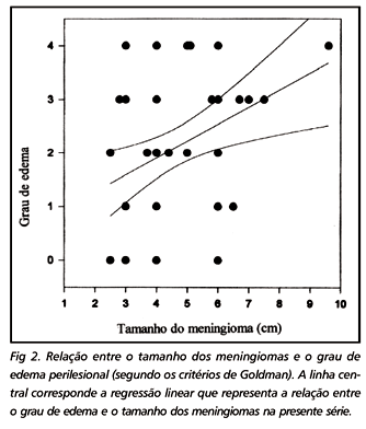
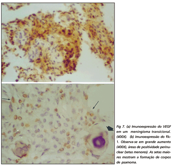
Many factors have been associated to the development of peritumoral edema in meningiomas. We studied the radiological and pathological features of 51 intracranial meningiomas surgically treated in the University Hospital of the Federal University of Rio de Janeiro. Two thirds of the meningiomas in our series had peritumoral edema. The size of the meningioma was probably related to the development of edema. Peritumoral edema was found frequently in the large meningiomas. The location of the meningioma was also associated with the frequency of peritumoral edema. Sphenoid wing meningiomas had significantly more peritumoral edema. In contrast, tuberculum sellae meningiomas were almost never associated with cerebral edema. There is growing evidence in the literature that vascular endothelial growth factor (VEGF) is a key factor in the pathogenesis of peritumoral edema. We studied by immunohistochemical techniques the expression of VEGF and its receptor flk-1 in meningiomas.
brain edema; immunohistochemistry; computerized tomography; magnetic resonance imaging


