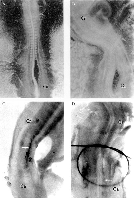
Important advances have been made in understanding the genetic processes that control skeletal muscle formation. Studies conducted on quails detected a delay in the myogenic program of animals selected for high growth rates. These studies have led to the hypothesis that a delay in myogenesis would allow somitic cells to proliferate longer and consequently increase the number of embryonic myoblasts. To test this hypothesis, recently segmented somites and part of the unsegmented paraxial mesoderm were separated from the neural tube/notochord complex in HH12 chicken embryos. In situ hybridization and competitive RT-PCR revealed that MyoD transcripts, which are responsible for myoblast determination, were absent in somites separated from neural tube/notochord (1.06 and 0.06 10-3 attomol MyoD/1 attomol ß-actin for control and separated somites, respectively; P<0.01). However, reapproximation of these structures allowed MyoD to be expressed in somites. Cellular proliferation was analyzed by immunohistochemical detection of incorporated BrdU, a thymidine analogue. A smaller but not significant (P = 0.27) number of proliferating cells was observed in somites that had been separated from neural tube/notochord (27 and 18 for control and separated somites, respectively). These results confirm the influence of the axial structures on MyoD activation but do not support the hypothesis that in the absence of MyoD transcripts the cellular proliferation would be maintained for a longer period of time.
Chicken development; MyoD; Myogenesis; In situ hybridization; BrdU; Cellular proliferation


