 i-motifs: DNA structures with regulatory functions and therapeutic potential
ABSTRACT
Non-B DNA structures represent alternative conformations to the canonical double helix, characterized by unconventional base pairing that diverge from the Watson-Crick model. Among these, the i-motifs, formed in cytosine-rich regions, has attracted considerable attention due to its potential regulatory roles. This review examines the molecular mechanisms underlying the formation and stability of i-motifs, which rely on hydrogen bonding between protonated cytosines. These structures can form under slightly acidic and even neutral pH conditions, depending on the DNA sequence context. Their diverse topologies and capacity to interact with specific proteins and ligands suggest that i-motifs play important roles in the regulation of gene expression, particularly within promoters of oncogenes, as well as in telomeric and centromeric regions. Understanding the behavior and biological relevance of i-motifs may deepen our insight into genomic regulation and reveal novel therapeutic opportunities. Targeting i-motif structures therefore represents a promising strategy for the treatment of cancer and other complex diseases.
Keywords:
Nucleic acid conformation; Molecular targeted therapy; Gene expression regulation; Oncogenes; Promoter regions; genetics
INTRODUCTION
The discovery of the DNA structure by Watson et al. in 1953 marked the beginning of a revolution in molecular biology. The double helix has since become the canonical model of genetic material, representing the mechanism by which biological information is stored and transmitted across generations. In this structure, the nitrogenous bases adenine and thymine are connected by two hydrogen bonds, whereas guanine and cytosine are linked by three.(1) However, the double helix does not encompass the full structural diversity of DNA. More recent studies have identified alternative conformations, known as non-B DNA structures, among which the i-motif has attracted significant attention.(2)
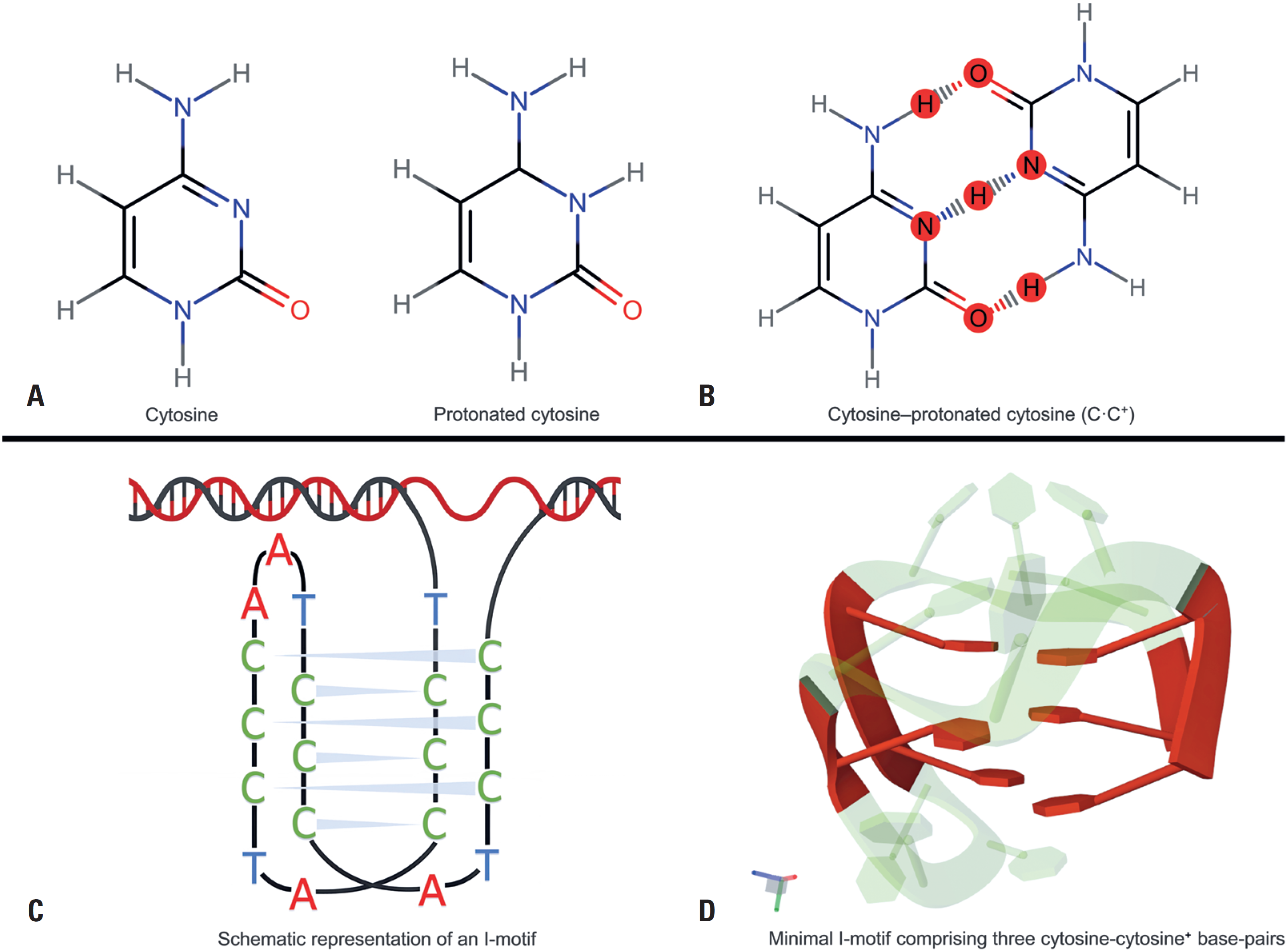
The i-motif is notable for its unconventional base-pairing pattern: instead of pairing with guanine, as is typical for cytosine, two cytosines interact directly, forming a hemiprotonated C·C+ base pair under specific conditions, typically at slightly acidic pH (Figure 1A-B).(3) This seemingly counter-intuitive phenomenon is increasingly recognized as part of a more versatile structural repertoire of DNA than previously appreciated. Interestingly, the term "i-motif" was used in scientific contexts even before its identification in human DNA, suggesting that this configuration may have implications beyond the human genome, including in fields such as virology.(4)
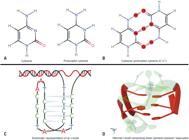
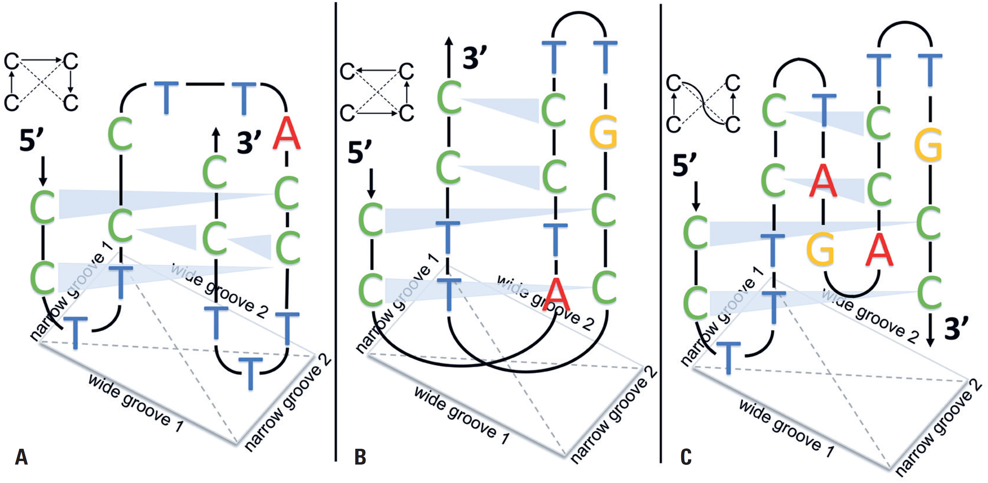
Protonated cytosine and C·C+ base pairing as the molecular basis of i-motif formation. (A) Chemical structures of neutral cytosine and protonated cytosine (C+), in which the additional proton is located at the N3 position. (B) Hemiprotonated cytosine–cytosine (C·C+) base pair. In this schematic, three hydrogen-bonding interactions are shown: one between the protonated N3–H+ and the N3 of the opposing base, and two involving the amino group (–NH2) and the carbonyl oxygen (C=O). Although three interactions are shown here for completeness, the formation of two hydrogen bonds (N3–H+···N3 and NH2–H···O=C) is generally sufficient and most commonly described in the literature as stabilizing the C·C+ base pair (adapted from Benabou et al., 2014).(11) (C) Schematic representation of the i-motif fold, highlighting the intercalated C·C+ base pairs that stabilize the structure. (D) Three-dimensional i-motif structure comprising three C·C+ base pairs (PDB ID: 8PMB), illustrating the characteristic tetrahelical fold (reported by Ghezzo et al., 2023).(12) The three-dimensional structure of the i-motif is relatively compact, consisting of four intercalated DNA strands arranged in an antiparallel orientation, with cytosine bases stacking to stabilize the structure. Each layer of hemiprotonated cytosines stacks upon the next, promoting additional interactions that maintain structural integrity. However, the i-motif can adopt different topologies - i.e., distinct three-dimensional patterns - depending on the nucleotide sequence (Figure 2). These base-stacking interactions are similar to those observed in other non-B structures, such as G-quadruplexes; however, i-motif stability specifically depends on cytosine protonation.(13) Van der Waals forces between stacked cytosine layers also contribute significantly to the structural stability
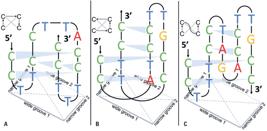
Schematic representation of different i-motif topologies, illustrating how variations in nucleotide sequence influence the spatial arrangement of four antiparallel DNA strands. (A) Basic organization of the i-motif, with stacked hemiprotonated cytosines base pairs (C·C+) forming the layers. The 5’ and 3’ ends are indicated, along with the "wide" and "narrow" clefts. (B) and (C) Alternative topologies, in which other bases (G, A, and T) are present in loops or at the termini, altering the overall conformation. Black arrows and curves illustrate strand connectivity, while shaded regions represent base stacking, contributing to structural stability. Adapted from Han et al., 1998(10)
The pH conditions that favor i-motif formation, ranging from slightly acidic to near-neutral, are not uncommon in animal cells.(5) This helps explain why its presence has been confirmed not only in humans but also in other mammals and even bacteria. (6,7) Techniques such as nuclear magnetic resonance, UV spectroscopy, and gel chromatography have been used to investigate its properties, revealing both the conditions that stabilize this structure and its potential biological functions.( Although the i-motif is currently one of the most extensively studied non-B DNA structures, it is not the only documented structural variation of DNA. G-quadruplexes, hairpins, cruciforms, and triplex structures are additional examples of non-B conformations that challenge traditional views of DNA's structural rigidity. The ability of DNA to adopt such diverse conformations not only underscores its complexity but also suggests that these variations may play functional roles that remain to be fully elucidated.
In recent years, - a growing body of research has focused on understanding how i-motifs may influence pathological processes, including cancer development. Although studies are still at an early stage, the possibilities are intriguing: these structures may be involved in gene regulatory mechanisms or in alterations that promote disease progression.(8,9) This perspective, while challenging, points to a fertile area of investigation that may redefine our understanding of the genome. Given the evidence presented, this narrative review aims to contextualize and integrate studies on non-B DNA structures, with a particular emphasis on i-motifs. This work synthesizes both theoretical and experimental contributions, highlighting how these structural variations and the molecular mechanisms governing them influence gene expression and are associated with pathological processes, such as cancer development. Additionally, it provides a historical overview of the discovery of these structures and traces the evolution of knowledge over time. By offering a comprehensive and up-to-date perspective on the subject, this review aims to promote innovative therapeutic approaches and stimulate further advances in the field of molecular biology.
Non-B structures
The i-motif is a secondary DNA structure that forms in cytosine-rich regions, typically under slightly acidic pH conditions. However, evidence suggests that specific sequences, such as d(5mCCT3CCT3ACCT3CC), can adopt the i-motif conformation even at neutral pH.(10) Unlike the conventional B-DNA helix, which relies on Watson-Crick hydrogen bonding between nitrogenous bases, the i-motif depends on unconventional interactions involving protonated cytosines. These interactions occur between two cytosines, one of which must be protonated (i.e., acquire an H+) to enable bond formation. The C+–C base pair is stabilized by hydrogen bonds: one between the amino group (NH2) of one cytosine and the carbonyl group (C=O) of the other, and another between the protonated N3–H+ and the N3 of the opposing base (Figure 1B).(11) i-motifs are commonly illustrated in figure 1, where a single DNA strand folds back on itself, forming the characteristic structure distinct from the canonical B-DNA conformation. The three-dimensional structure of the i-motif is highly compact, consisting of four intercalated strands arranged in an antiparallel orientation, with cytosine bases stacking to stabilize the structure (Figure 1D).(12)
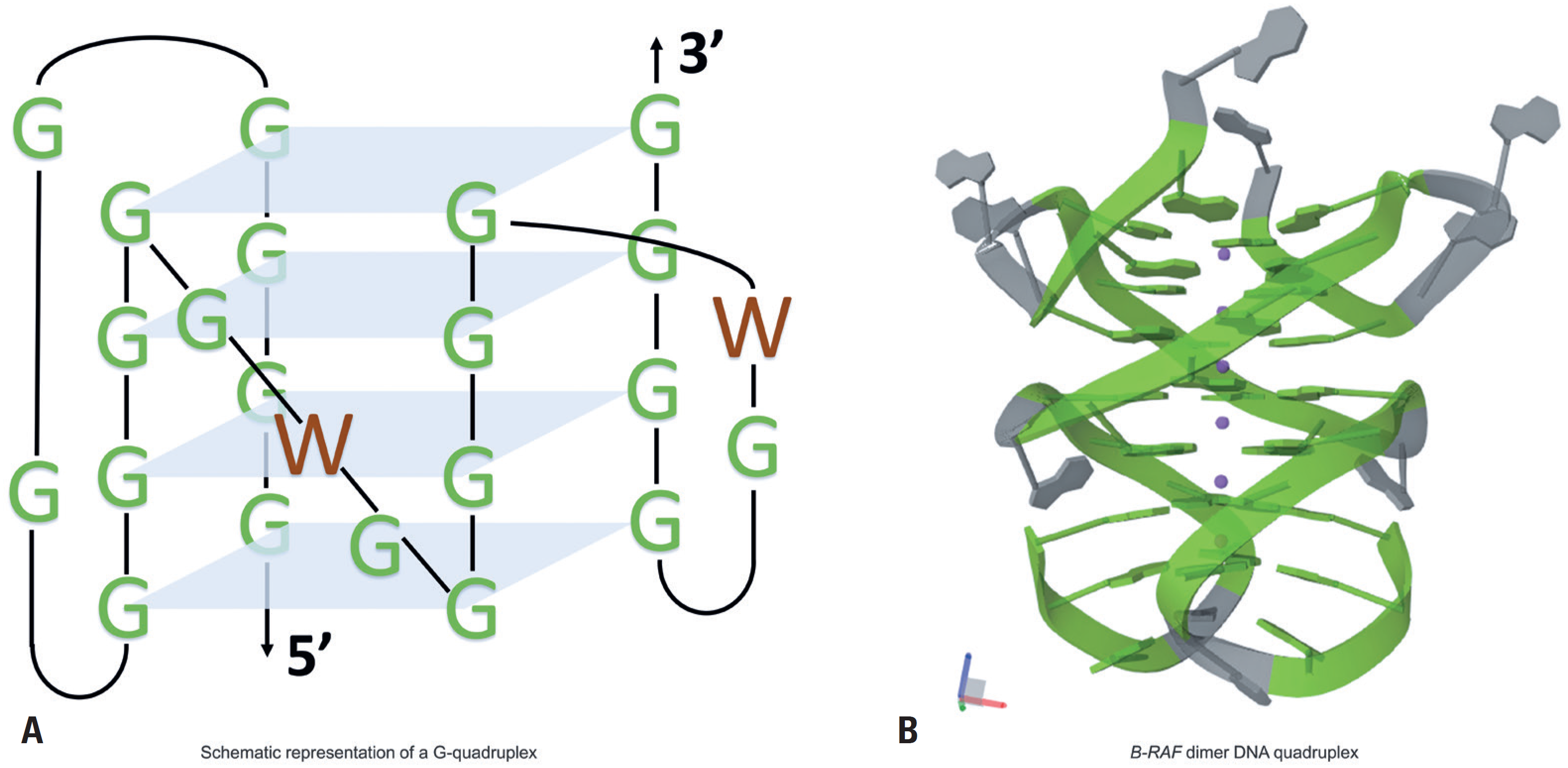
G-quadruplexes (G4s) are four-stranded DNA structures that form in guanine-rich regions.(14) These structures are stabilized by Hoogsteen hydrogen bonding, which differs from canonical Watson-Crick base pairing. Each G4 consists of four guanine bases arranged in a planar configuration, forming a guanine tetrad stabilized by monovalent cations such as potassium (K+) or sodium (Na+), which are positioned between the tetrads to enhance stability.(15) Depending on sequence length and strand orientation, G4s can adopt parallel, antiparallel, or hybrid conformations. Specific G4 topologies have been associated with particular genomic regions and pathological features, such as in the PDGFA gene, which has been linked to metastatic potential (Figure 3).(16)
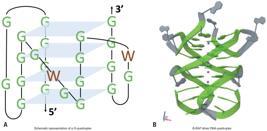
G-quadruplex DNA. (A) Guanine bases are stabilized by Hoogsteen hydrogen bonding; the letter "W" marks positions that can engage in canonical Watson–Crick base pairing. Adapted from Choi & Majima, 2011.(2) (B) Atomic model of the human BRAF promoter G-quadruplex (PDB 4H29; X-ray), which adopts an intertwined dimeric arrangement. Guanine residues are shown in green and form the G-quadruplex; while other nucleotides are shown in gray. Potassium ions in the central channel are depicted as purple spheres (structure from Wei et al., 2013)(14)
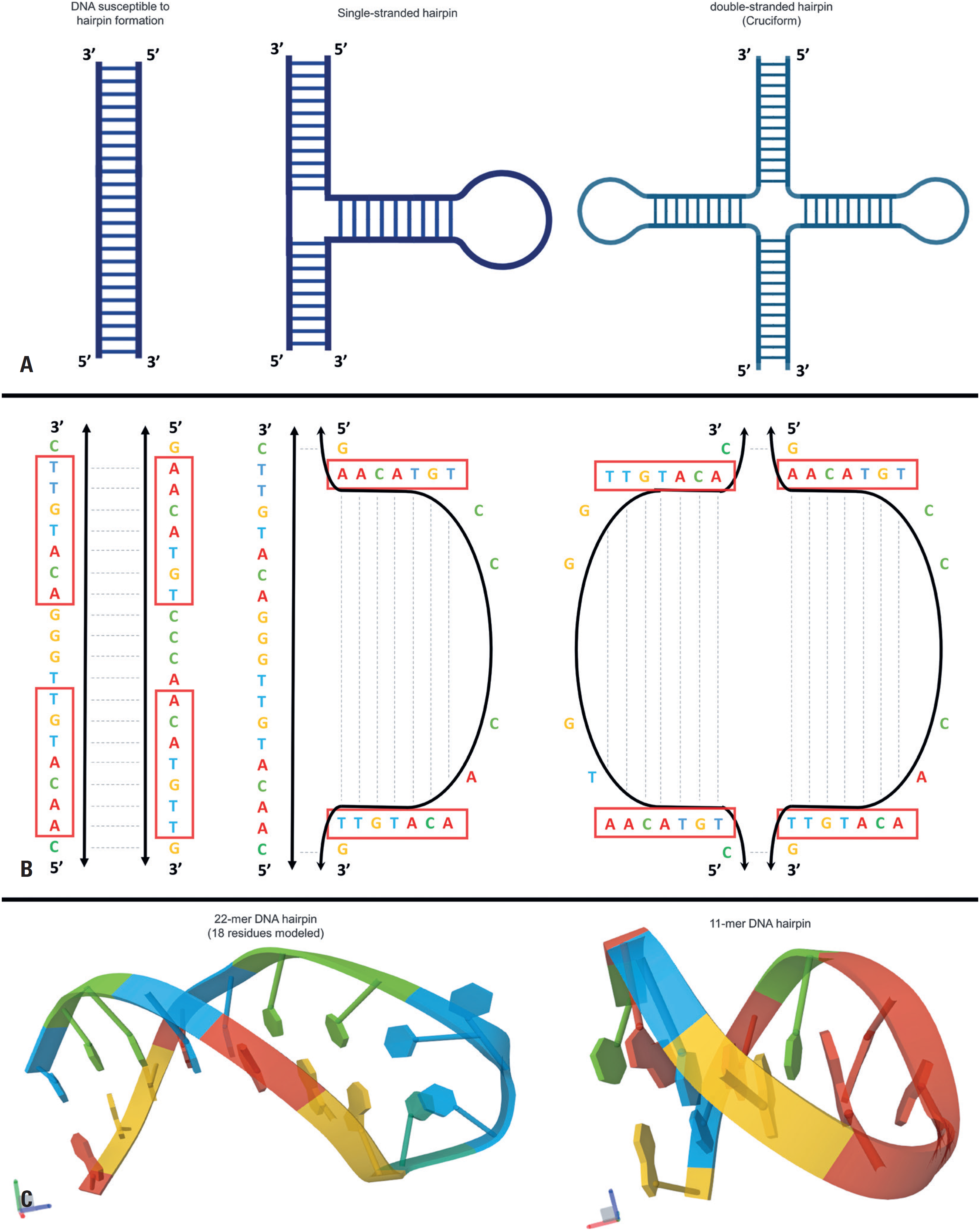
Hairpins and cruciforms are structures formed when a single DNA or RNA strand folds back on itself, creating a paired "stem" of complementary bases and an unpaired "loop." These structures commonly arise in inverted repeat sequences, where intramolecular base pairing can occur. When two such hairpin structures form on opposite strands of DNA, they can give rise to a more complex structure known as a cruciform. These structures may interfere with replication and transcription by acting as physical barriers that stall the enzymes involved in these processes. Regions of negative supercoiling favor cruciform formation and are often associated with palindromic repeat sequences (Figure 4).(17
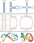
DNA hairpins and cruciforms. (A) Schematic overview. Left: a duplex segment containing an inverted repeat sequence (a short palindromic motif) that is susceptible to hairpin extrusion. Middle: a single-stranded hairpin formed by intramolecular base pairing. Right: a cruciform structure formed when both strands of the inverted repeat fold out to generate two opposing hairpins. (B) Sequence-level view of hairpin formation. Palindromic segments that pair to form the stem are boxed; dashed lines denote Watson–Crick base pairs; arrows indicate the 5′→3′ orientation; adapted from Bansal et al., 2022.(17) (C) Representative three-dimensional hairpin structures from the RCSB Protein Data Bank. Left: an 18-nt (18-nucleotide) hairpin (PDB ID: 1QE7, solution NMR; Ghosh et al., 1999).(18) Right: an 11-nt hairpin (PDB ID 1BJH, solution NMR; Chou et al., 1996).(19) An atomic-resolution three-dimensional cruciform structure is not shown because, to our knowledge, such structures have not been deposited in the PDB as stable, isolated entries. Cruciform formation depends on DNA topology and negative supercoiling and is often transient in short oligonucleotides, making it difficult to capture using NMR or crystallography
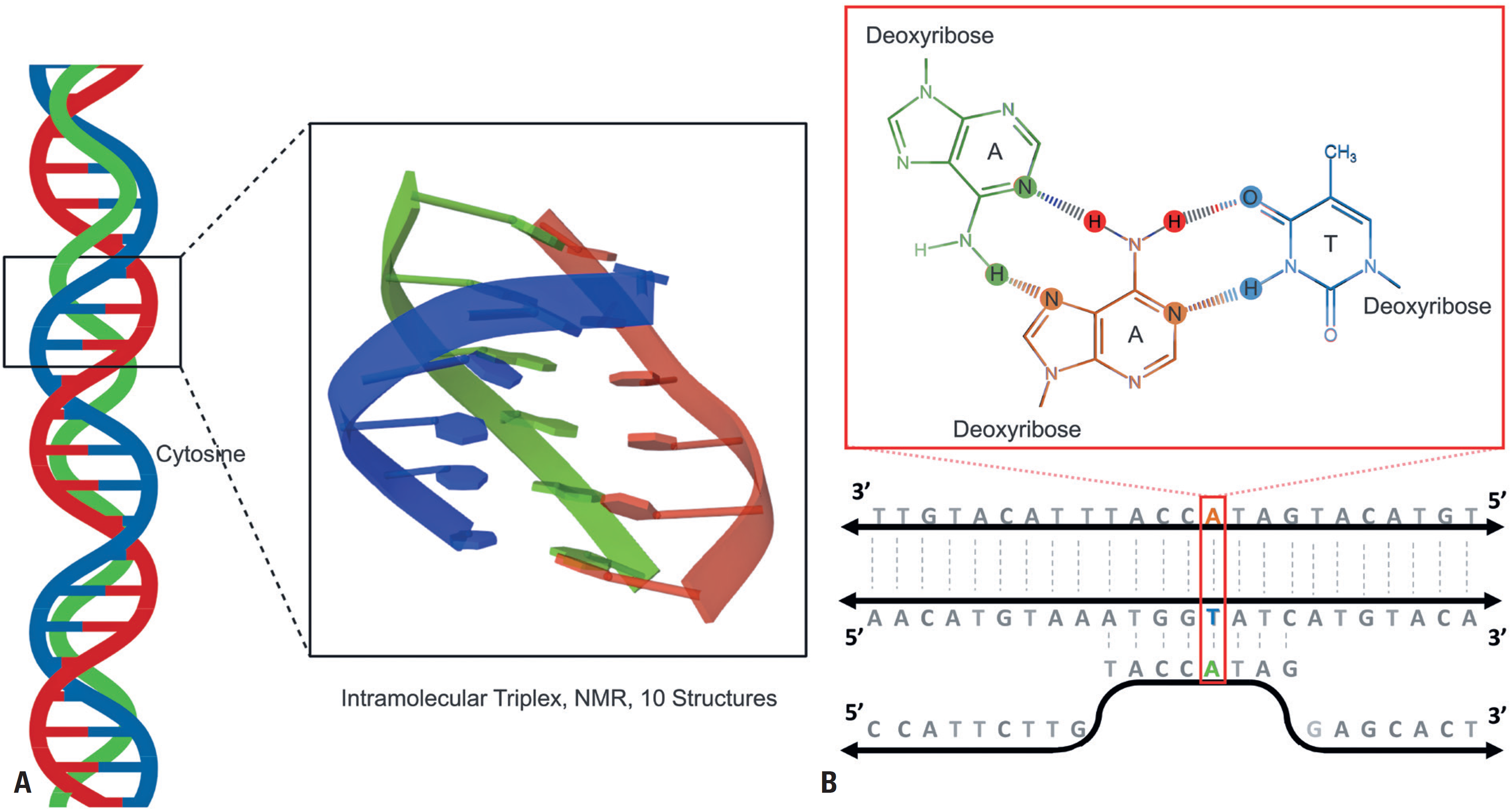
Triplex structures, or triple helices, form when a third DNA strand binds to the canonical double helix. This third strand may originate from a separate DNA molecule or from a different region of the same molecule, folding back to interact with the double helix (Figure 5).(20) The third strand binds to the double helix through Hoogsteen or reverse Hoogsteen hydrogen bonding, which differs from canonical Watson-Crick base pairing.(21) In Watson-Crick base pairing, adenine (A) pairs with thymine (T) via two hydrogen bonds, while guanine (G) pairs with cytosine (C) via three hydrogen bonds. In Hoogsteen bonding, the base-pairing geometry is altered, allowing a third base to interact with purines (A or G) in one strand of the double helix. Purines, due to their larger size and two-ring structure (compared to the single-ring structure of pyrimidines), are more accessible for these additional interactions.(2,22,23) This three-stranded structure can interfere with critical processes such as DNA replication and transcription
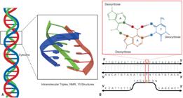
DNA triple helix (triplex) structures. A) Three-dimensional view of an intramolecular DNA triplex solved by solution-state nuclear magnetic resonance (PDB ID: 1BCB). The structure was deposited by Asensio et al. (1998) and classified as DNA without mutations.(20) This triple helical arrangement illustrates the association of a third strand with the canonical duplex, stabilized by Hoogsteen interactions. B) Simplified schematic (adapted from Brazda et al., 2020; and Holder et al., 2015) illustrating the principle of triplex formation.(22,23) The canonical duplex is stabilized by Watson–Crick base pairs, while the third strand binds in the major groove via Hoogsteen hydrogen bonds. The chemical structure shown in green corresponds to adenine from the third strand, forming a representative T·A·T triad with a Watson–Crick A·T base pair. Such alternative base-pairing interactions enable an additional strand to associate with duplex DNA through Hoogsteen hydrogen bonding, generating a triple-helical structure capable of modulating essential processes such as replication and transcription
Triplex structures may be recognized as DNA damage by repair proteins such as ERCC1-XPF and XPG. These proteins, which typically detect and repair genuine DNA lesions, may identify triple helices as damaged and attempt to process them. During this process, they may inadvertently introduce DNA breaks, thereby increasing genomic instability and the risk of mutations and chromosomal rearrangements.(21)
Biological functions of the i-motif structure
The i-motif is a secondary DNA structure characterized as a four-stranded non-covalent complex that forms in cytosine-rich sequences, typically under slightly acidic pH conditions.(2) Unlike the canonical DNA double helix (B-DNA), i-motifs have been less extensively studied than other non-B structures, such as G-quadruplexes. However, interest in these structures has grown significantly in recent years, driven by their potential regulatory roles in biological processes, particularly in the modulation of gene expression. Their biological functions are closely linked to their ability to interact with various molecules, including proteins, small ligands, and epigenetic modifications.(24,25)
I-motifs are proposed to act as molecular "on-off" switches for gene expression, influencing essential cellular processes such as replication, transcription, and translation.(26,27) These structures are found in several regulatory regions of the human genome, including promoters, telomeres, centromeres, and ribosomal DNA regions.(24,26) Their presence in gene promoters, for example, underscores their structural diversity, including variations in topology, loop length, and transition pH. This versatility makes them promising targets for investigating their biological functions and for the development of small-molecule-based therapies.(26,27)
The interaction of i-motifs with other molecules is crucial for their biological functions, although the precise mechanisms underlying gene expression regulation are not yet fully understood. Evidence suggests that i-motifs interact with DNA-binding proteins, serving as recognition sites for transcription factors or repressors.(28-30) These interactions may modulate gene expression by either facilitating or hindering the binding of regulatory proteins. In some cases, i-motifs physically impede transcription factor binding, thereby repressing gene expression.(26)
On the other hand, specific ligands may induce conformational changes in the i-motif, thereby increasing its affinity for regulatory proteins and leading to transcriptional activation and enhanced gene expression.(26,27) The interaction of i-motifs with proteins such as hnRNP LL and hnRNP K has been extensively studied. For instance, hnRNP LL interacts with i-motifs, promoting their unfolding and the formation of a stable single-stranded structure, which facilitates transcriptional activation.(31,32) Similarly, hnRNP K has been shown to unfold the i-motif in the KRAS gene region, thereby positively regulating its expression. Additionally, i-motifs formation may compete with other DNA structures, such as G-quadruplexes, which are also involved in gene regulation.(33) The transition between these conformations may be modulated by environmental factors, such as pH and ionic strength, providing a dynamic mechanism for the control of gene expression.(15,26) The influence of ligands on i-motif-mediated gene expression is complex and multifaceted. Ligands may modulate i-motif stability, either stabilizing or destabilizing the structure. In some cases, stabilization may prevent transcription factor binding, leading to gene repression, or impede RNA polymerase progression, thereby inhibiting transcription.(24,34) A notable example is the natural compound hyoscine butylbromide, which specifically binds to the G-quadruplex within the C-MYB gene.(35) Furthermore, ligands may influence epigenetic processes, such as DNA methylation, thereby indirectly affecting gene expression.(36-38) Compounds such as IMC-48 and IMC-76 have been shown to modulate gene expression by interacting with i-motif-forming sequences in the BCL2 promoter, exerting opposite effects on BCL2 regulation.(26,39) Another example is the stabilization of telomeric i-motifs by carboxylated single-walled carbon nanotubes (SWCNTs), which inhibit telomerase activity and interfere with telomere functions in cancer cells.(40)
The stability of i-motifs under physiological conditions is a critical factor for their biological relevance. Although i-motifs were initially thought to be stable only at acidic pH, recent studies have that some can form at neutral pH, particularly in sequences containing at least five consecutive cytosines.(40) In addition to pH, factors such as low temperature, molecular crowding,(13) negative supercoiling,(28-30) and the presence of silver (I) cations also influence i-motif stability.(29) A notable example is the i-motif-forming sequence in the promoter region of HIF-1α, which exhibits stability at near-neutral pH.(5)
In the therapeutic context, i-motifs are frequently found in oncogene promoter regions and may be considered complementary to G-quadruplexes.(26) Given the role of G-quadruplexes in telomerase inhibition, transcriptional regulation, and replication, it is plausible that i-motifs also exert regulatory functions, either as primary elements or by contributing to the stabilization of G-quadruplexes relative to the double helix. This versatility makes i-motifs promising targets for chemical intervention, particularly in contexts in which they may directly modulate transcription.(40) A relevant example is the stabilization of telomeric i-motifs by carboxylated single-walled carbon nanotubes, which inhibit telomerase activity and interfere with telomere function in cancer cells.(41)
In conclusion, i-motifs are non-canonical DNA structures that appear to play significant roles in biological processes, particularly in the regulation of gene expression. Although research on i-motifs is still at an early stage, current evidence highlights their potential as therapeutic targets for a variety of diseases. Further studies are needed to fully elucidate their biological functions and to develop small molecules capable of specifically modulating these structures for therapeutic purposes.
Implications of i-motif structures in carcinogenesis
Cytosines and guanine-rich regions are frequently found in oncogene promoters, centromeres, and telomeres. These regions have the potential to form specific non-canonical DNA structures, such as G-quadruplexes and i-motifs.(28,42)
Since i-motifs are formed by cytosines, nucleotides that represent the primary targets of epigenetic modifications in the human genome, they serve as potential sites for epigenetic regulation. These modifications can directly influence crucial biological processes(38) as changes at the 5-position of cytosines within CpG dinucleotides affect the thermal stability of non-canonical structures, particularly i-motifs.(25,43)
In promoter regions, i-motifs may act as molecular regulators, modulating gene expression and contributing to genome integrity.(36) Their abundance increases during the G1/S phase of the cell cycle, a period marked by intense transcriptional activity. This association highlights a possible link between i-motifs and cancer development, as these structures are thought to function as recognition sites for proteins involved in transcriptional activation.(44) Furthermore, i-motifs have been identified in the promoters of several cancer-related genes, including HRAS,(32) KRAS,(45) VEGFA,(46) RET,(47) BCL2,(48) RAD17,(49) MYC,(50) MYB,(51) KIT,(52) PDGFR-B,(53) HIF-1A,(54) RB1,(55) and TERT (Table 1).(56)
Relationship between cancer-related genes, their functions, and i-motif-mediated transcriptional modulation: summary of gene functions and i-motif-associated transcriptional regulation, highlighting evidence reported in the scientific literature
The presence of i-motif structures in the promoter regions of oncogenes, as mentioned earlier, does not directly alter gene transcription. However, their stability and conformation can be modulated by proteins or small molecules. These interactions may lead to either activation or repression of gene expression, highlighting the dynamic regulatory role of these structures.
Although i-motifs are recognized as regulatory elements, demonstrating their functionality in vivo remains a significant challenge. These structures are highly dynamic and transient, making them difficult to detect under physiological conditions. While early studies suggested that i-motifs were stable only in acidic environments, more recent work has shown that specific sequences, such as those in HIF-1A and telomeric regions, fold at near neutral pH.(13,40) However, such stability is context-dependent, and factors such as negative supercoiling, molecular crowding, and protein interactions are crucial for their formation. This complexity has fueled ongoing debate as to whether i-motifs function as regulatory switches or merely represent transient structural intermediates.
Although i-motifs have been identified in numerous genes, as summarized in table 1, the functional relationship between i-motif formation and gene transcription remains unclear for many of them. This highlights a substantial gap in our understanding of their biological significance.
Another limitation lies in the methodological constraints of current detection tools. Traditional biophysical approaches, such as circular dichroism and NMR spectroscopy, are often performed under non-physiological conditions and cannot fully capture the dynamics of i-motif folding in living cells. Although emerging strategies, such as fluorescent probes, i-motif–specific antibodies, and in-cell NMR spectroscopy, have provided compelling evidence of their presence in vivo, these techniques still face challenges related to sensitivity, specificity, and spatial resolution.(13)
i-motifs in centromeres and telomeres: implications for chromosomal stability
Centromeres, the chromosomal regions where kinetochores (multiprotein structures responsible for attaching chromosomes to spindle microtubules) are assembled, are essential for proper chromosome segregation and sister chromatid separation during cell division.(71,72) Alterations in centromeric stability are closely associated with tumor development.(72) Studies have identified i-motif structures in centromeric satellite DNA, and their presence has been positively associated with the structural and functional organization of centromeres.(73,74) Evidence suggests that defects in centromere formation and maintenance may precede tumor cell development, as disruptions in these domains have been frequently observed across various cancer types.(75,76) This suggests a potential protective role of i-motifs in limiting tumor initiation and progression.
Complementarily, telomeres– nucleoprotein complexes located at chromosome ends that prevent inappropriate DNA repair responses, contain guanine- and cytosine-rich sequences capable of adopting G-quadruplex and i-motif conformations.(15,77) Telomere stability is regulated by the enzyme telomerase, whose activity is crucial in carcinogenesis, as telomere maintenance is a common feature of various tumor cell types.(78) The formation of G-quadruplex and i-motif structures at telomeres has been associated with telomerase inhibition, leading to telomere uncapping and subsequent activation of a DNA damage response, which may ultimately result in growth arrest and compromise cancer cell survival.(41,79)
Implications of i-motifs in pathological conditions beyond cancer
I-motif mediated gene regulation presents promising applications in pathological conditions beyond cancer. In hepatic disorders such as non-alcoholic steatohepatitis (NASH), stabilization of the i-motif in the BCL-2 gene has been explored as an innovative therapeutic strategy. The use of A22, an acridone derivative, demonstrated significant efficacy in stabilizing the i-motif located in the BCL2 promoter, resulting in increased transcription and reduced apoptosis in hepatocytes. In animal models, this approach concurrently improved metabolic and inflammatory markers, suggesting a systemic impact rarely observed with single-target therapeutic strategies.(8)
The investigation into the relationship between i-motifs and other diseases has also expanded to neuropsychiatric conditions. Thorne et al. examined this association in the context of neuropsychiatric disorders. They demonstrated that the SLC6A4 gene, responsible for encoding the serotonin transporter, contains allelic variants (VNTR-S and VNTR-L) that modulate transporter expression and influence susceptibility to psychiatric disorders. These variants are enriched in cytosine-guanine-rich regions, suggesting the potential formation of i-motifs. In vitro spectroscopy confirmed the folding of these regions into i-motif structures, indicating that such conformations may also occur in vivo, potentially regulating SLC6A4 expression.(80)
Another relevant study on i-motifs, this time in the context of viral infections, was conducted by Ruggiero et al. The authors demonstrated that the HIV genome contains a region known as the long terminal repeat (LTR), which is essential for viral replication following integration into the human genome. This region is enriched in cytosine- and guanine- rich sequences capable of forming i-motifs, whose stabilization inhibits LTR transcription and consequently viral replication. Based on this premise, the study investigated the hnRNP K protein as a stabilizer of the i-motif within this region, enhancing its resistance to variations in pH and temperature. Stabilization of i-motif structures in the LTR showed potential to reduce viral replication, representing an innovative strategy for the development of antiretroviral therapies targeting this region.(81)
Beyond their role in gene regulation, i-motifs have also been explored as functional components for controlled drug delivery. Hu et al. developed a DNA hydrogel capable of carrying insulin while resisting the acidic environment of the stomach and subsequently releasing the drug in the intestine. The hydrogel's structure, enriched in hemiprotonated cytosine bases that favor i-motif formation, provided stability to the system until it reached the intestinal tract. The results demonstrated the potential of this approach for drug administration in diabetes treatment, ensuring controlled release throughout the gastrointestinal tract.(82)
CONCLUSION
Recent advances have significantly expanded our understanding of i-motif DNA structures, positioning them as dynamic elements with potential regulatory and therapeutic implications. Although traditionally regarded as transient intermediates, studies published over the past five years have shown that certain i-motifs can form and remain stable under near-physiological conditions, particularly in regulatory regions such as promoters, telomeres, and centromeres. These developments have been driven by the advent of high-resolution imaging, real-time detection systems, and structure-specific probes, most notably the use of i-motif, specific antibodies, and in-cell NMR, which have provided unprecedented insight into their dynamics in living cells.
Despite these advances, key questions remain unanswered, including whether i-motifs exert causal regulatory roles or act primarily as structural intermediates, how their formation is orchestrated during the cell cycle, and to what extent they contribute to genome stability and disease pathogenesis. Addressing these gaps will require integrated strategies combining genome editing (e.g., CRISPR/Cas), structure-targeting ligands, and multi-omics approaches to map their biological functions with greater resolution and context specificity.
Future research should also prioritize functional validation in model organisms and leverage single-molecule biophysics to link i-motif folding dynamics to transcriptional control. Such studies will be crucial for determining whether i-motifs are viable therapeutic targets and for translating these findings into innovative interventions.
In conclusion, i-motifs represent structurally unique and biologically significant DNA elements, offering a fertile field for discovery at the interface of structural biology, epigenetics, and translational medicine. A deeper mechanistic understanding will be essential to fully realize their potential as biomarkers and therapeutic targets in cancer and other complex diseases.
-
AUTHORS’ STATEMENT ON GENERATIVE ARTIFICIAL INTELLIGENCE
The authors declare that generative Artificial Intelligence tools were not used to write or generate scientific content in this manuscript. Grammarly for Windows, v.1.2.253.1650, was used solely for grammar and language refinement and did not affect the scientific content, data interpretation, or conclusions.
DATA AVAILABILITY
The underlying content is contained within the manuscript.
REFERENCES
- 1 Watson JD, Crick FH. Molecular structure of nucleic acids; a structure for deoxyribose nucleic acid. Nature. 1953;171(4356):737-8.
- 2 Choi J, Majima T. Conformational changes of non-B DNA. Chem Soc Rev. 2011;40(12):5893-909.
- 3 Gehring K, Leroy JL, Guéron M. A tetrameric DNA structure with protonated cytosine.cytosine base pairs. Nature. 1993;363(6429):561-5.
- 4 Xiao JH, Davidson I, Macchi M, Rosales R, Vigneron M, Staub A, et al. In vitro binding of several cell-specific and ubiquitous nuclear proteins to the GT-I motif of the SV40 enhancer. Genes Dev. 1987;1(8):794-807.
- 5 Dzatko S, Krafcikova M, Hänsel-Hertsch R, Fessl T, Fiala R, Loja T, et al. Evaluation of the Stability of DNA i-Motifs in the Nuclei of Living Mammalian Cells. Angew Chem Int Ed Engl. 2018;57(8):2165-9.
- 6 Serrano-Chacón I, Mir B, Escaja N, González C. Structure of i-Motif/Duplex Junctions at Neutral pH. J Am Chem Soc. 2021;143(33):12919-23.
- 7 Leroy JL, Guéron M, Mergny JL, Hélène C. Intramolecular folding of a fragment of the cytosine-rich strand of telomeric DNA into an i-motif. Nucleic Acids Res. 1994;22(9):1600-6.
- 8 Li X, Wang J, Gong X, Zhang M, Kang S, Shu B, et al. Upregulation of BCL-2 by acridone derivative through gene promoter i-motif for alleviating liver damage of NAFLD/NASH. Nucleic Acids Res. 2020;48(15):8255-68.
- 9 Li KS, Jordan D, Lin LY, McCarthy SE, Schneekloth JS Jr, Yatsunyk LA. Crystal Structure of an i-Motif from the HRAS Oncogene Promoter. Angew Chem Int Ed Engl. 2023;62(26):e202301666.
- 10 Han X, Leroy JL, Guéron M. An intramolecular i-motif: the solution structure and base-pair opening kinetics of d(5mCCT3CCT3ACCT3CC). J Mol Biol. 1998;278(5):949-65.
- 11 Benabou S, Aviñó A, Eritja R, González C, Gargallo R. Fundamental aspects of the nucleic acid i-motif structures. RSC Adv. 2014;4(51):26956-80.
- 12 Ghezzo M, Trajkovski M, Plavec J, Sissi C. A Screening Protocol for Exploring Loop Length Requirements for the Formation of a Three Cytosine-Cytosine+ Base-Paired i-Motif. Angew Chem Int Ed Engl. 2023;62(41):e202309327.
- 13 Zeraati M, Langley DB, Schofield P, Moye AL, Rouet R, Hughes WE, et al. I-motif DNA structures are formed in the nuclei of human cells. Nat Chem. 2018;10(6):631-7.
- 14 Wei D, Todd AK, Zloh M, Gunaratnam M, Parkinson GN, Neidle S. Crystal structure of a promoter sequence in the B-raf gene reveals an intertwined dimer quadruplex. J Am Chem Soc. 2013;135(51):19319-29.
- 15 Wolski P, Nieszporek K, Panczyk T. G-Quadruplex and I-Motif Structures within the Telomeric DNA Duplex. A Molecular Dynamics Analysis of Protonation States as Factors Affecting Their Stability. J Phys Chem B. 2019;123(2):468-79.
- 16 Brooks TA, Kendrick S, Hurley L. Making sense of G-quadruplex and i-motif functions in oncogene promoters. FEBS J. 2010;277(17):3459-69.
- 17 Bansal A, Kaushik S, Kukreti S. Non-canonical DNA structures: diversity and disease association. Front Genet. 2022;13:959258.
- 18 Ghosh M, Kumar NV, Varshney U, Chary KV. Structural characterisation of a uracil containing hairpin DNA by NMR and molecular dynamics. Nucleic Acids Res. 1999;27(19):3938-44.
- 19 Chou SH, Zhu L, Gao Z, Cheng JW, Reid BR. Hairpin loops consisting of single adenine residues closed by sheared A.A and G.G pairs formed by the DNA triplets AAA and GAG: solution structure of the d(GTACAAAGTAC) hairpin. J Mol Biol. 1996;264(5):981-1001.
- 20 Asensio JL, Brown T, Lane AN. Comparison of the solution structures of intramolecular DNA triple helices containing adjacent and non-adjacent CG.C+ triplets. Nucleic Acids Res. 1998;26(16):3677-86.
- 21 Jain A, Wang G, Vasquez KM. DNA triple helices: biological consequences and therapeutic potential. Biochimie. 2008;90(8):1117-30.
- 22 Brazda V, Fojta M, Bowater RP. Structures and stability of simple DNA repeats from bacteria. Biochem J. 2020;477(2):325-39.
- 23 Holder IT, Wagner S, Xiong P, Sinn M, Frickey T, Meyer A, et al. Intrastrand triplex DNA repeats in bacteria: a source of genomic instability. Nucleic Acids Res. 2015;43(21):10126-42.
- 24 Luo X, Zhang J, Gao Y, Pan W, Yang Y, Li X, et al. Emerging roles of i-motif in gene expression and disease treatment. Front Pharmacol. 2023;14:1136251.
- 25 Oshikawa D, Inaba S, Kitagawa Y, Tsukakoshi K, Ikebukuro K. CpG Methylation Altered the Stability and Structure of the i-Motifs Located in the CpG Islands. Int J Mol Sci. 2022;23(12):6467.
- 26 Sedghi Masoud S, Nagasawa K. i-Motif-Binding Ligands and Their Effects on the Structure and Biological Functions of i-Motif. Chem Pharm Bull (Tokyo). 2018;66(12):1091-103.
- 27 Das S, Takahashi S, Ohyama T, Bhowmik S, Sugimoto N. Theranostic approach to specifically targeting the interloop region of BCL2 i-motif DNA by crystal violet. Sci Rep. 2023;13(1):14338.
- 28 Brazier JA, Shah A, Brown GD. I-motif formation in gene promoters: unusually stable formation in sequences complementary to known G-quadruplexes. Chem Commun (Camb). 2012;48(87):10739-41.
- 29 Day HA, Huguin C, Waller ZA. Silver cations fold i-motif at neutral pH. Chem Commun (Camb). 2013;49(70):7696-8.
- 30 Sun D, Hurley LH. The importance of negative superhelicity in inducing the formation of G-quadruplex and i-motif structures in the c-Myc promoter: implications for drug targeting and control of gene expression. J Med Chem. 2009;52(9):2863-74.
- 31 McLuckie KI, Waller ZA, Sanders DA, Alves D, Rodriguez R, Dash J, et al. G-quadruplex-binding benzo[a]phenoxazines down-regulate c-KIT expression in human gastric carcinoma cells. J Am Chem Soc. 2011;133(8):2658-63.
- 32 Miglietta G, Cogoi S, Pedersen EB, Xodo LE. GC-elements controlling HRAS transcription form i-motif structures unfolded by heterogeneous ribonucleoprotein particle A1. Sci Rep. 2015;5(1):18097.
- 33 Bhavsar-Jog YP, Van Dornshuld E, Brooks TA, Tschumper GS, Wadkins RM. Epigenetic modification, dehydration, and molecular crowding effects on the thermodynamics of i-motif structure formation from C-rich DNA. Biochemistry. 2014;53(10):1586-94.
- 34 Galburt EA, Grill SW, Wiedmann A, Lubkowska L, Choy J, Nogales E, et al. Backtracking determines the force sensitivity of RNAP II in a factor-dependent manner. Nature. 2007;446(7137):820-3.
- 35 Wang S, Yang Y, Fan S, Li H, David DY. [Determination of the binding of natural products to the human c-myb oncogene promoter G-quadruplex DNA by capillary electrophoresis and electrospray ionization mass spectrometry]. Se Pu. 2020;38(9):1069-77.
- 36 Školáková P, Badri Z, Foldynová-Trantírková S, Ryneš J, Šponer J, Fojtová M, et al. Composite 5-methylations of cytosines modulate i-motif stability in a sequence-specific manner: Implications for DNA nanotechnology and epigenetic regulation of plant telomeric DNA. Biochim Biophys Acta Gen Subj. 2020;1864(9):129651.
- 37 Wright EP, Abdelhamid MA, Ehiabor MO, Grigg MC, Irving K, Smith NM, et al. Epigenetic modification of cytosines fine tunes the stability of i-motif DNA. Nucleic Acids Res. 2020;48(1):55-62.
- 38 Xu B, Devi G, Shao F. Regulation of telomeric i-motif stability by 5-methylcytosine and 5-hydroxymethylcytosine modification. Org Biomol Chem. 2015;13(20):5646-51.
- 39 Kendrick S, Kang HJ, Alam MP, Madathil MM, Agrawal P, Gokhale V, et al. The dynamic character of the BCL2 promoter i-motif provides a mechanism for modulation of gene expression by compounds that bind selectively to the alternative DNA hairpin structure. J Am Chem Soc. 2014;136(11):4161-71.
- 40 Wright EP, Huppert JL, Waller ZA. Identification of multiple genomic DNA sequences which form i-motif structures at neutral pH. Nucleic Acids Res. 2017;45(6):2951-9.
- 41 Chen Y, Qu K, Zhao C, Wu L, Ren J, Wang J, et al. Insights into the biomedical effects of carboxylated single-wall carbon nanotubes on telomerase and telomeres. Nat Commun. 2012;3(1):1074.
- 42 Garavís M, González C, Villasante A. On the origin of the eukaryotic chromosome: the role of noncanonical DNA structures in telomere evolution. Genome Biol Evol. 2013;5(6):1142-50.
- 43 Kimura K, Oshikawa D, Ikebukuro K, Yoshida W. Stabilization of VEGF i-motif structure by CpG methylation. Biochem Biophys Res Commun. 2022;594:88-92.
- 44 Abou Assi H, Garavís M, González C, Damha MJ. i-Motif DNA: structural features and significance to cell biology. Nucleic Acids Res. 2018;46(16):8038-56.
- 45 Kaiser CE, Van Ert NA, Agrawal P, Chawla R, Yang D, Hurley LH. Insight into the Complexity of the i-Motif and G-Quadruplex DNA Structures Formed in the KRAS Promoter and Subsequent Drug-Induced Gene Repression. J Am Chem Soc. 2017;139(25):8522-36.
- 46 Uribe DJ, Guo K, Shin YJ, Sun D. Heterogeneous nuclear ribonucleoprotein K and nucleolin as transcriptional activators of the vascular endothelial growth factor promoter through interaction with secondary DNA structures. Biochemistry. 2011;50(18):3796-806.
- 47 Guo K, Pourpak A, Beetz-Rogers K, Gokhale V, Sun D, Hurley LH. Formation of pseudosymmetrical G-quadruplex and i-motif structures in the proximal promoter region of the RET oncogene. J Am Chem Soc. 2007;129(33):10220-8.
- 48 Kang HJ, Kendrick S, Hecht SM, Hurley LH. The transcriptional complex between the BCL2 i-motif and hnRNP LL is a molecular switch for control of gene expression that can be modulated by small molecules. J Am Chem Soc. 2014;136(11):4172-85.
- 49 Rogers RA, Fleming AM, Burrows CJ. Unusual Isothermal Hysteresis in DNA i-Motif pH Transitions: A Study of the RAD17 Promoter Sequence. Biophys J. 2018;114(8):1804-15.
- 50 Dai J, Hatzakis E, Hurley LH, Yang D. I-motif structures formed in the human c-MYC promoter are highly dynamic--insights into sequence redundancy and I-motif stability. PLoS One. 2010;5(7):e11647.
- 51 Li H, Hai J, Zhou J, Yuan G. The formation and characteristics of the i-motif structure within the promoter of the c-myb proto-oncogene. J Photochem Photobiol B. 2016;162:625-32.
- 52 Gong X, Lin X, Wang S, Ji D, Shu B, Huang ZS, et al. Regulation of c-Kit gene transcription selectively by bisacridine derivative through promoter dual i-motif structures. Biochim Biophys Acta Gene Regul Mech. 2023;1866(2):194912.
- 53 Brown RV, Wang T, Chappeta VR, Wu G, Onel B, Chawla R, et al. The consequences of overlapping G-quadruplexes and i-motifs in the platelet-derived growth factor receptor β core promoter nuclease hypersensitive element can explain the unexpected effects of mutations and provide opportunities for selective targeting of both structures by small molecules to downregulate gene expression. J Am Chem Soc. 2017;139(22):7456-75.
- 54 Cheng M, Chen J, Ju H, Zhou J, Mergny JL. Drivers of i-DNA Formation in a Variety of Environments Revealed by Four-Dimensional UV Melting and Annealing. J Am Chem Soc. 2021;143(20):7792-807.
- 55 Lin X, Zhang J, Liang J, Ji D, Huang ZS, Li D. Selective Up-Regulation of Tumor Suppressor Gene Retinoblastoma by Bisacridine Derivative Through Gene Promoter Quadruplex Structures for Cancer Treatment. Int J Mol Sci. 2025;26(4):1417.
- 56 Conrad JW, Sowers ML, Yap DY, Cherryhomes E, Pettitt BM, Khanipov K, et al. Transition Mutations in the hTERT Promoter Are Unrelated to Potential i-motif Formation in the C-Rich Strand. Biomolecules. 2023;13(9):1308.
- 57 Simanshu DK, Nissley DV, McCormick F. RAS Proteins and Their Regulators in Human Disease. Cell. 2017;170(1):17-33.
- 58 Ferrara N. The role of vascular endothelial growth factor in pathological angiogenesis. Breast Cancer Res Treat. 1995;36(2):127-37.
- 59 Manié S, Santoro M, Fusco A, Billaud M. The RET receptor: function in development and dysfunction in congenital malformation. Trends Genet. 2001;17(10):580-9.
- 60 Vaux DL, Cory S, Adams JM. Bcl-2 gene promotes haemopoietic cell survival and cooperates with c-myc to immortalize pre-B cells. Nature. 1988;335(6189):440-2.
- 61 Shinohara M, Sakai K, Ogawa T, Shinohara A. The mitotic DNA damage checkpoint proteins Rad17 and Rad24 are required for repair of double-strand breaks during meiosis in yeast. Genetics. 2003;164(3):855-65.
- 62 Shim H, Dolde C, Lewis BC, Wu CS, Dang G, Jungmann RA, et al. c-Myc transactivation of LDH-A: implications for tumor metabolism and growth. Proc Natl Acad Sci USA. 1997;94(13):6658-63.
- 63 Tomonaga T, Levens D. Activating transcription from single stranded DNA. Proc Natl Acad Sci USA. 1996;93(12):5830-5.
- 64 Ramsay RG, Barton AL, Gonda TJ. Targeting c-Myb expression in human disease. Expert Opin Ther Targets. 2003;7(2):235-48.
- 65 Shin JY, Hu W, Naramura M, Park CY. High c-Kit expression identifies hematopoietic stem cells with impaired self-renewal and megakaryocytic bias. J Exp Med. 2014;211(2):217-31.
- 66 Colucci F, Di Santo JP. The receptor tyrosine kinase c-kit provides a critical signal for survival, expansion, and maturation of mouse natural killer cells. Blood. 2000;95(3):984-91.
- 67 Wu E, Palmer N, Tian Z, Moseman AP, Galdzicki M, Wang X, et al. Comprehensive dissection of PDGF-PDGFR signaling pathways in PDGFR genetically defined cells. PLoS One. 2008;3(11):e3794.
- 68 Weidemann A, Johnson RS. Biology of HIF-1α. Cell Death Differ. 2008;15(4):621-7.
- 69 Dyson NJ. RB1: a prototype tumor suppressor and an enigma. Genes Dev. 2016;30(13):1492-502.
- 70 Yuan X, Larsson C, Xu D. Mechanisms underlying the activation of TERT transcription and telomerase activity in human cancer: old actors and new players. Oncogene. 2019;38(34):6172-83.
- 71 Garavís M, Escaja N, Gabelica V, Villasante A, González C. Centromeric Alpha-Satellite DNA Adopts Dimeric i-Motif Structures Capped by AT Hoogsteen Base Pairs. Chemistry. 2015;21(27):9816-24.
- 72 Nassar R, Thompson L, Fouquerel E. Molecular mechanisms protecting centromeres from self-sabotage and implications for cancer therapy. NAR Cancer. 2023;5(2):zcad019.
- 73 Garavís M, Méndez-Lago M, Gabelica V, Whitehead SL, González C, Villasante A. The structure of an endogenous Drosophila centromere reveals the prevalence of tandemly repeated sequences able to form i-motifs. Sci Rep. 2015;5(1):13307.
- 74 Nonin-Lecomte S, Leroy JL. Structure of a C-rich strand fragment of the human centromeric satellite III: a pH-dependent intercalation topology. J Mol Biol. 2001;309(2):491-506.
- 75 Knutsen T, Padilla-Nash HM, Wangsa D, Barenboim-Stapleton L, Camps J, McNeil N, et al. Definitive molecular cytogenetic characterization of 15 colorectal cancer cell lines. Genes Chromosomes Cancer. 2010;49(3):204-23.
- 76 Mackinnon RN, Campbell LJ. The role of dicentric chromosome formation and secondary centromere deletion in the evolution of myeloid malignancy. Genet Res Int. 2011;2011:643628.
- 77 Hänsel R, Löhr F, Foldynová-Trantírková S, Bamberg E, Trantírek L, Dötsch V. The parallel G-quadruplex structure of vertebrate telomeric repeat sequences is not the preferred folding topology under physiological conditions. Nucleic Acids Res. 2011;39(13):5768-75.
- 78 Shay JW, Bacchetti S. A survey of telomerase activity in human cancer. Eur J Cancer. 1997;33(5):787-91.
- 79 Neidle S, Parkinson GN. The structure of telomeric DNA. Curr Opin Struct Biol. 2003;13(3):275-83.
- 80 Thorne BN, Ellenbroek BA, Day DJ. Evaluation of i-Motif Formation in the Serotonin Transporter-Linked Polymorphic Region. Chembiochem. 2021;22(2):349-53.
- 81 Ruggiero E, Lago S, Šket P, Nadai M, Frasson I, Plavec J, et al. A dynamic i-motif with a duplex stem-loop in the long terminal repeat promoter of the HIV-1 proviral genome modulates viral transcription. Nucleic Acids Res. 2019;47(21):11057-68.
- 82 Hu Y, Gao S, Lu H, Ying JY. Acid-Resistant and Physiological pH-Responsive DNA Hydrogel Composed of A-Motif and i-Motif toward Oral Insulin Delivery. J Am Chem Soc. 2022;144(12):5461-70.
Edited by
-
Associate Editor:
Luciana Cavalheiro Marti Instituto Israelita de Ensino e Pesquisa Albert Einstein, São Paulo, SP, Brazil ORCID: https://orcid.org/0000-0002-3890-0827