 Development of a low-fidelity simulation model for ultrasound-guided vascular puncture
ABSTRACT
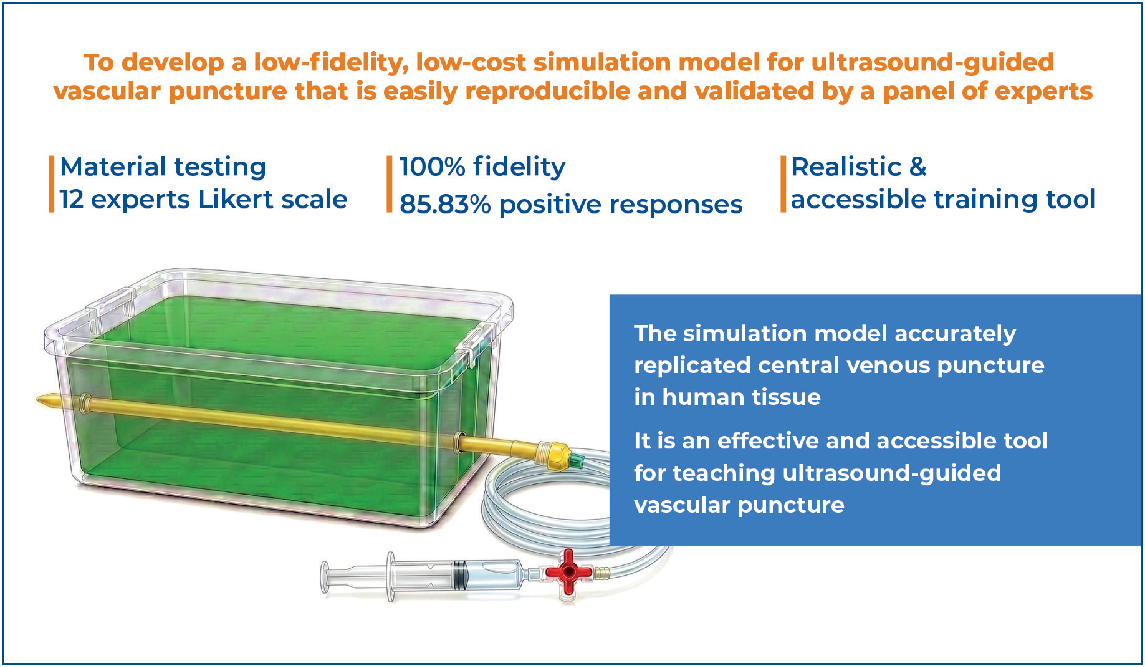
Objective: To develop a low-fidelity, low-cost simulation model for ultrasound-guided vascular puncture that is easily reproducible and validated by an expert panel.
Methods: Sequential tests using different materials were conducted based on phantom models described in the literature. The final model underwent evaluation by 12 volunteer expert physicians, who assessed usability, fidelity, and reproducibility during repeated ultrasound-guided punctures. Feedback was collected using a Likert scale instrument.
Results: All specialists agreed on model fidelity, including echogenicity, scenario reproduction, and tactile sensation, as well as suitability for training and reproducibility. Overall, positive responses accounted for 85.83% of evaluated items.
Conclusion: The proposed simulation model closely resembled the scenario of central venous puncture in human tissue and represented an effective and accessible tool for teaching ultrasound-guided vascular puncture techniques.
Keywords:
Central venous access; Ultrasonography, interventional; Ultrasonography; Education, medical; Simulation training; Phantoms, imaging
Highlights
-
■ Ultrasound improves success rates and reduces complications during vascular puncture.
-
■ Simulation-based training enables safe and effective acquisition of procedural skills.
-
■ A low-cost, reproducible simulation model was developed.
-
■ Specialists validated the model as effective for teaching and training.
In Brief
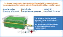
This study developed and validated a low-fidelity, low-cost simulation model for ultrasound-guided vascular puncture. Evaluation by specialists demonstrated adequate fidelity, usability, and reproducibility, indicating that the model is an effective and accessible teaching and training tool for the technique.
INTRODUCTION
Central venous access (CVA) can be performed using anatomical landmark techniques or ultrasound-guided approaches. The ultrasound-guided approach improves patient safety by enabling visualization of anatomical variations, thrombi, and stenoses and by allowing real-time verification of puncture and catheter placement.(1) Moreover, simulation contributes to the development of skills and competencies through realistic scenarios that support clinical reasoning and the application of procedural techniques.(2) These scenarios enhance patient safety by refining techniques through repetition, improving problem-solving, and enabling the identification and correction of errors committed by professionals compared with purely theoretical education.(3) Studies have reported reductions in mechanical complications, a 12% increase in first-puncture success, and a 72% decrease in inadvertent arterial puncture.(4,5) Consequently, several guidelines over the past few years have recommended ultrasound-guided CVA, with the most robust statistical evidence for the internal jugular vein.(5-14) However, simulation-based training for operating physicians remains essential for the effective application of this technique.
Simulation enables the development of skills and competencies in a controlled and protected environment without posing risk to patients,(15) while also improving technique and shortening the learning curve.(16) In this context, simulation promotes the motor coordination required for simultaneous needle puncture and transducer manipulation.(17) Training can be conducted using low-, medium-, or high-fidelity simulators. Although high-complexity simulators are commercially available, studies indicate that, in educational settings, the level of training fidelity is not necessarily associated with simulator realism, as other factors such as scenario design also influence performance.(18,19)
For ultrasound-guided puncture simulation, training phantoms(20) are available and may be synthetic or biological, as well as homemade or commercial. One limitation of commercial models is their high cost, which hinders widespread adoption.(21) Homemade phantoms provide a cost-effective alternative that offers adaptability and allows repetitive practice.(22) In an effort to promote ultrasound-guided CVA, this article aims to describe the construction of a homemade phantom with a favorable cost–benefit ratio that is easily reproducible, simple to store, suitable for repeated puncture, and ultrasonographically similar to human tissue, validated by a panel of experts.
OBJECTIVE
This study aims to develop a low-fidelity, low-cost simulation model for ultrasound-guided vascular puncture that is easily reproducible and validated by a panel of experts.
METHODS
Puncture model
A literature search was first conducted using the keywords "Phantom AND venous access" and "Homemade AND vascular access." In the first stage, materials were tested as substitutes for blood vessels, evaluating diameter, tactile sensation, ease of puncture, and compatibility with ultrasound imaging.
In the second stage, the composition of the phantom was developed based on three models previously reported in the literature. The criteria assessed included model echogenicity, tactile sensation during puncture, needle visualization, presence of artifacts, and storage stability outside refrigeration. Based on the tested and selected compositions for construction of the CVA puncture phantom, model reproducibility was assessed by two medical students at different times. They received instructions regarding the production steps, and after fabrication, the models were evaluated using ultrasound.
Validation by experts
A panel of experts was selected using convenience sampling based on the following criteria: residence in the project city, a specialty registered with the professional council, performance of ultrasound-guided punctures on a monthly basis, and at least five years of experience in ultrasound use. The specialists tested the developed phantom and completed a 14-item checklist using a Likert scale. The checklist includes four questions addressing the specialist's experience with ultrasound-guided punctures, while the remaining items evaluate the proposed model. These items were based on similar studies assessing homemade phantom models for training physicians in ultrasound-guided puncture.(17,23,24) The questions were adapted to reflect the specific characteristics intended to be addressed by the developed model as well as model usability, reliability, and reproducibility during serial ultrasound-guided punctures.
Statistical analysis
Data analysis was performed using non-parametric measures, including the median and interquartile range. Differences between group medians (surgeons versus non-surgeons) were tested using the Mann-Whitney–Wilcoxon test. To assess the degree of agreement among specialists, Gwet's AC2 agreement test was performed.(25-27) Analyses were conducted using the R statistical environment with a quantitative approach. The agreement coefficient was evaluated at a 5% significance level, with 95% confidence intervals reported.
RESULTS
Phase 1 – Development of a puncture model
Model structure: A tubular balloon demonstrated the best overall performance for vessel simulation after diferent materials tests. To accommodate the phantom, a disposable polypropylene container measuring 15.5cm in length, 10.5cm in width, and 5.5cm in height was used. Regarding the vessel simulator, seven different materials were selected and assessed for compatibility with human vessel diameters, tactile feedback during puncture, puncture success rate, and ultrasound image quality. The results are summarized in table 1.
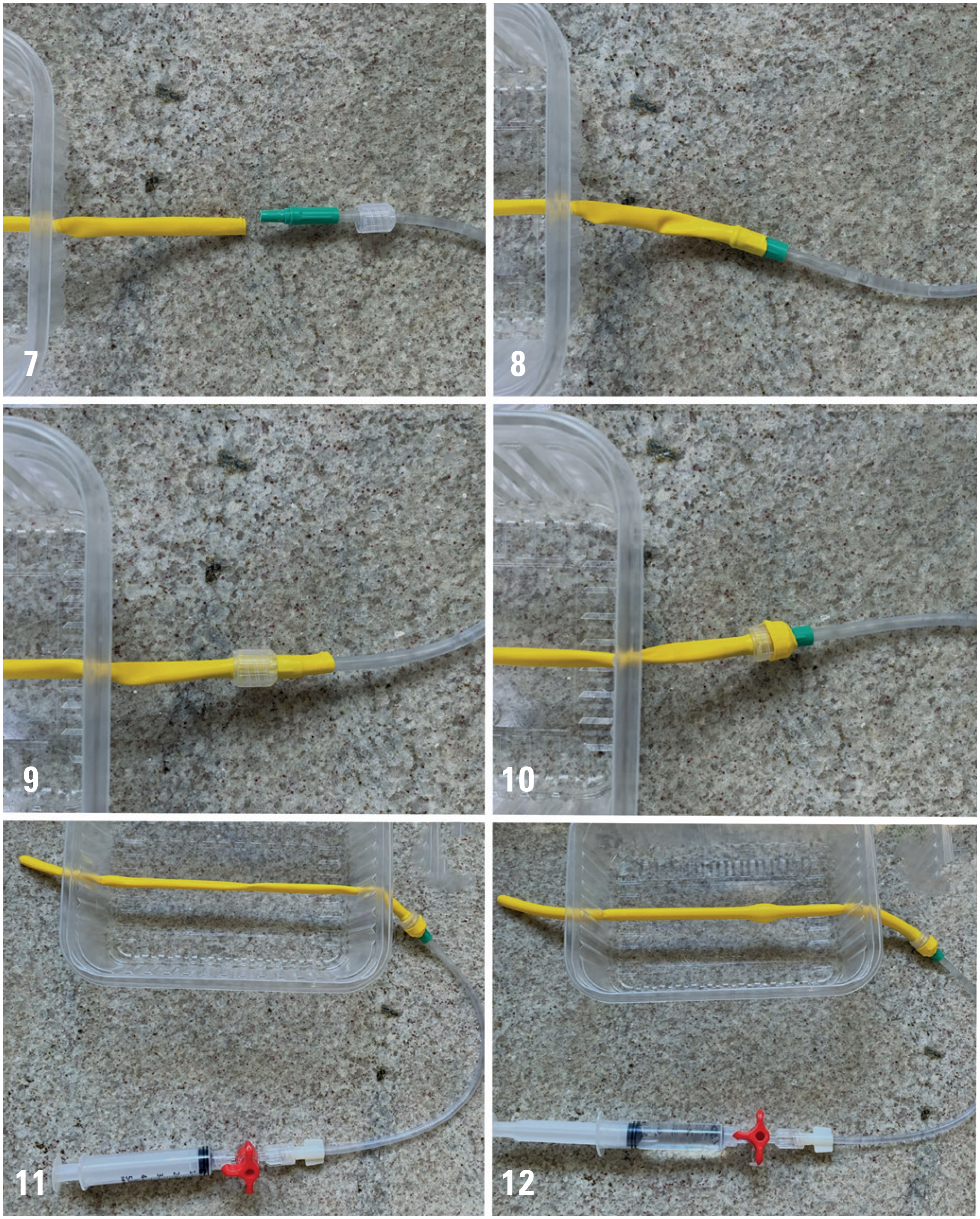
Following testing, the material that best fulfilled the criteria was the tubular balloon. The balloon was positioned inside the container through two lateral openings created with a heated metal object and then filled with water. To prevent deflation, a 20cm Luer-lock extension set, a three-way stopcock, and a 10mL syringe were attached.
Composition
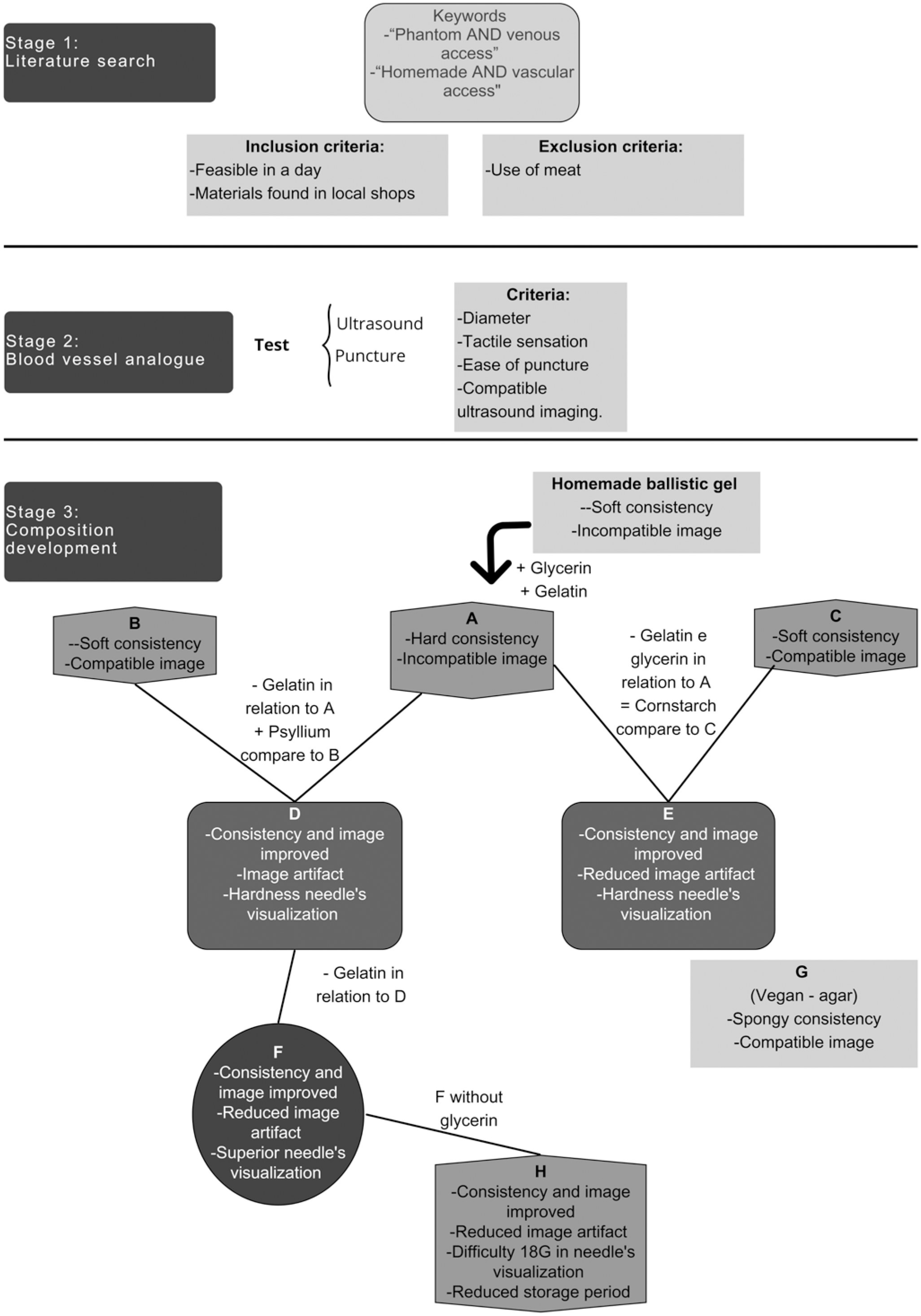
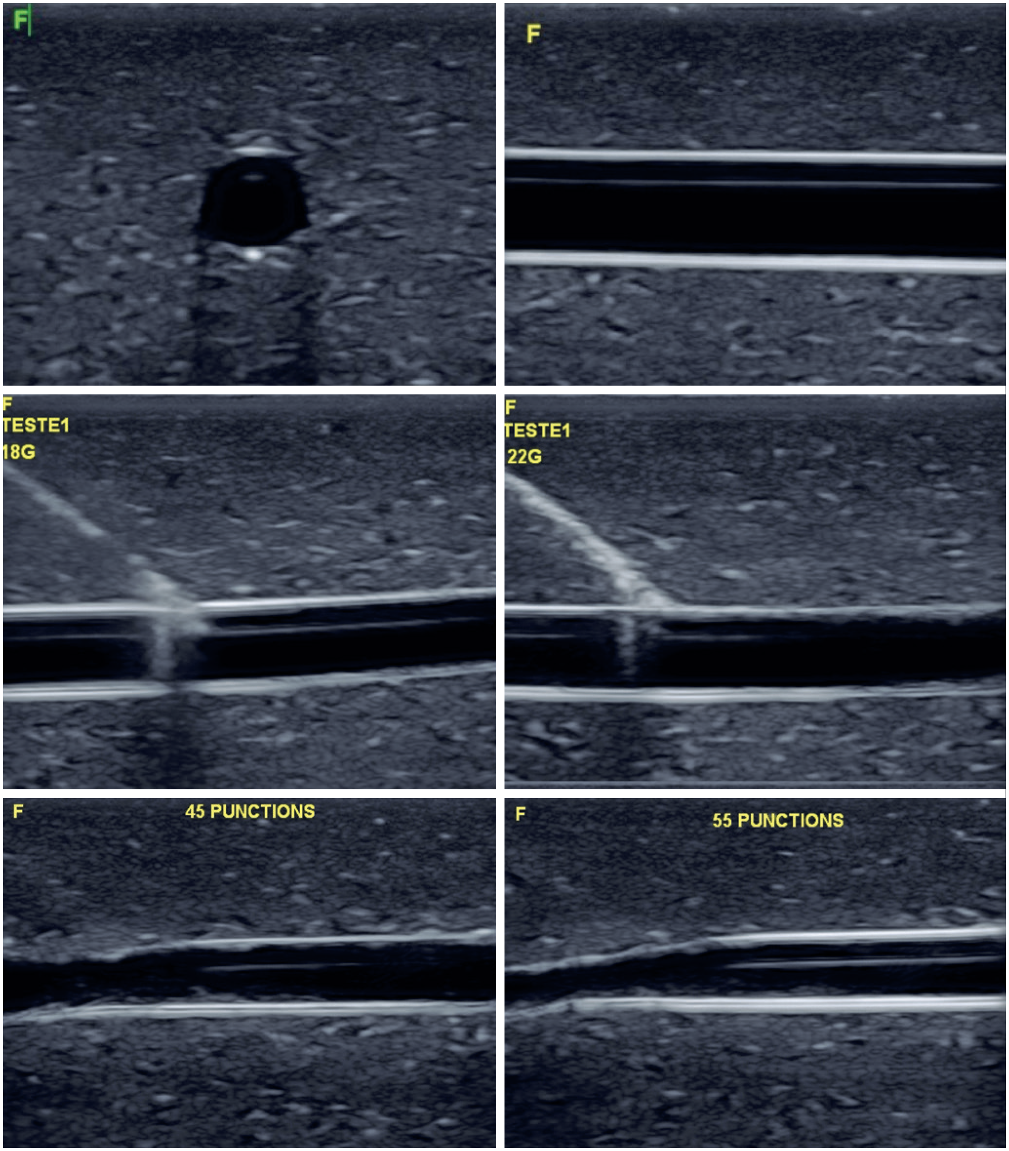
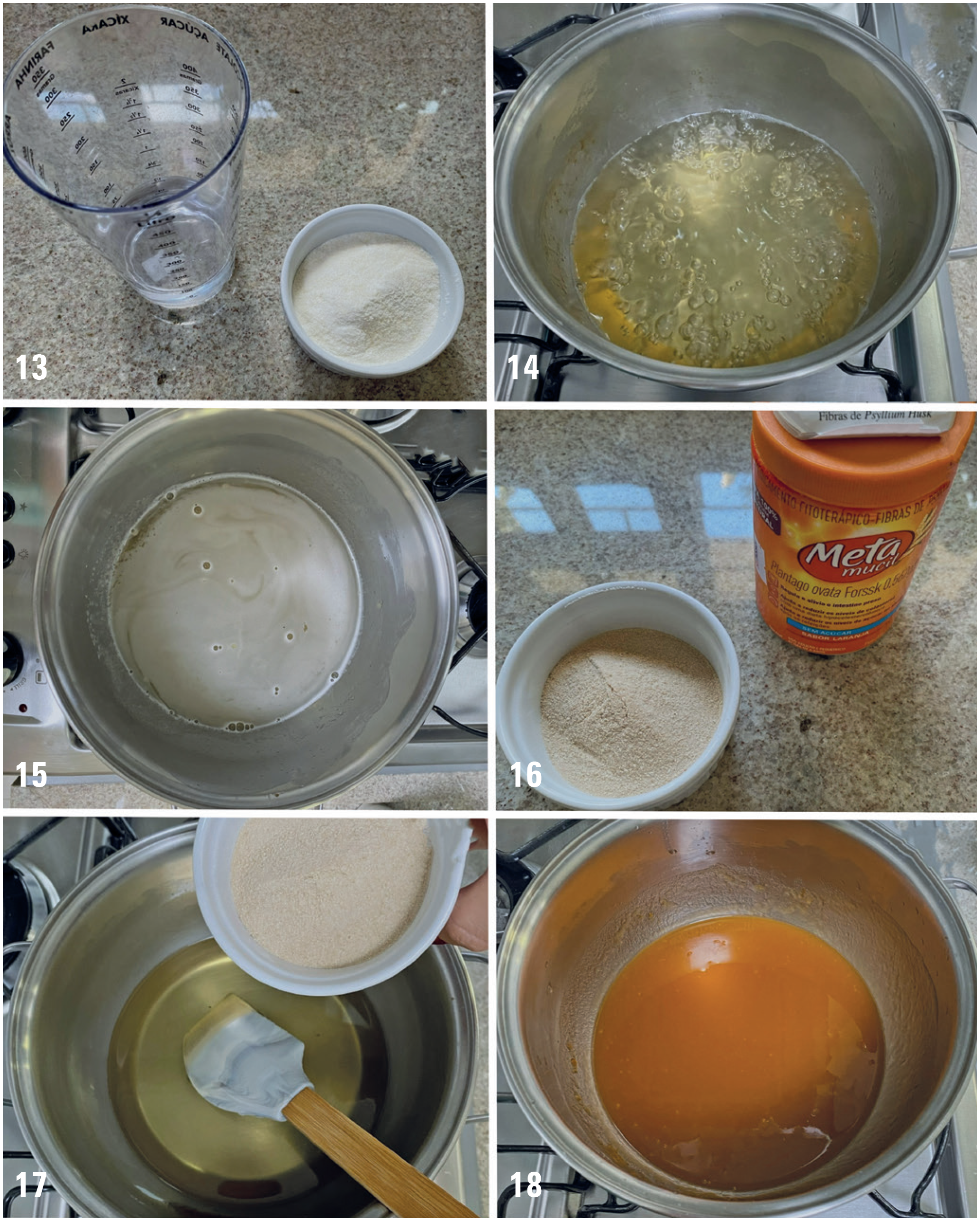
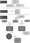
Nine formulations were testd and composition F best met the predefined criteria for the model. The formulation of the homemade phantom was based on recipes available in scientific databases, using ballistic gelatin(28-30) as the main component, along with a composition containing psyllium(23) and another in which corn flour(31) was replaced with cornstarch. Different recipes were prepared (Table 2), varying both the quantity and combination of ingredients (Figure 1), until the initial objective was achieved. At the end of the experiments, Composition F satisfied all the established criteria (Table 3 and Figure 2).
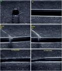
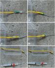
Ultrasound images of Composition F showing transverse and longitudinal views of the simulated vessel, needle puncture using 18G and 22G needles, and model integrity after 45 and 55 punctures
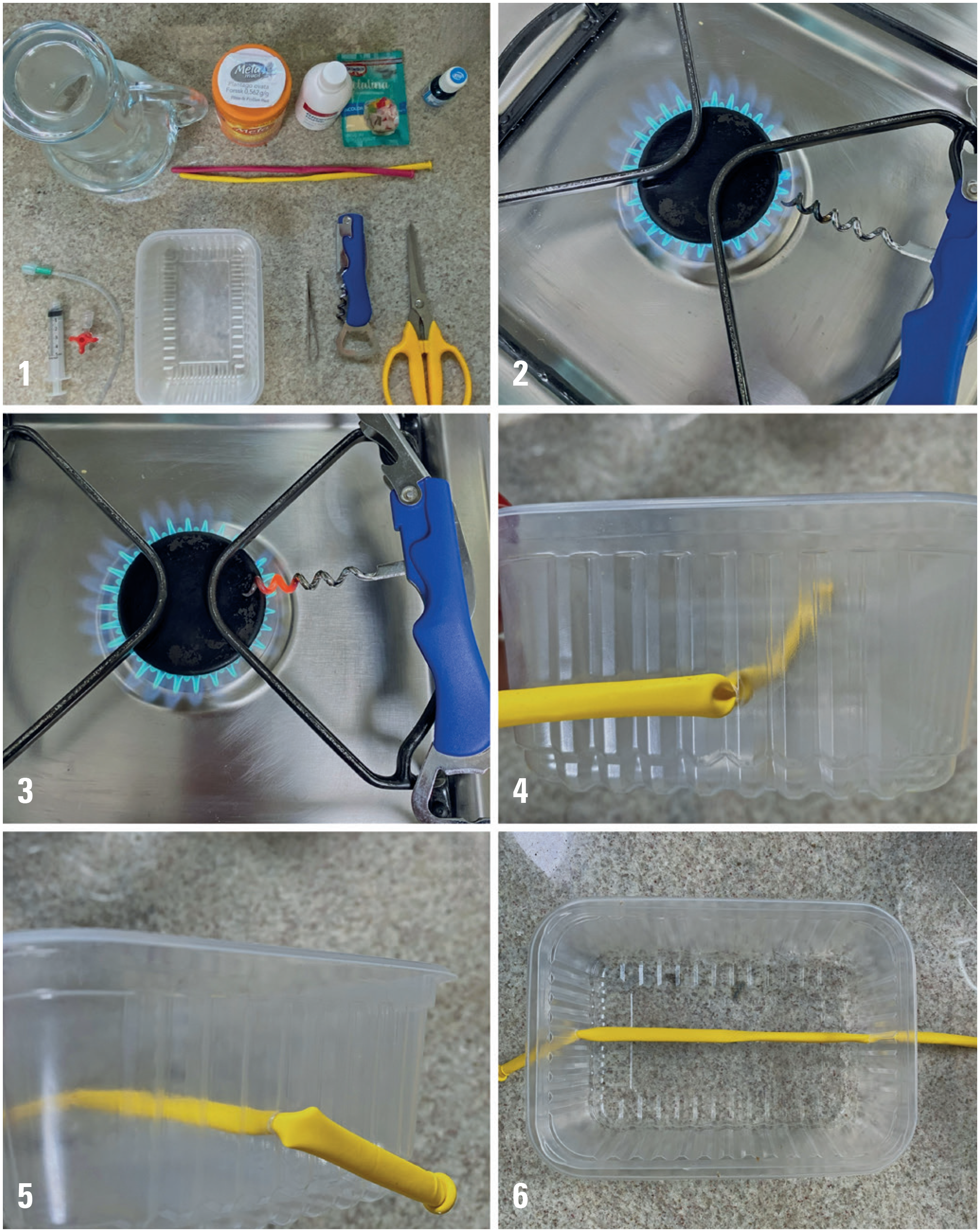
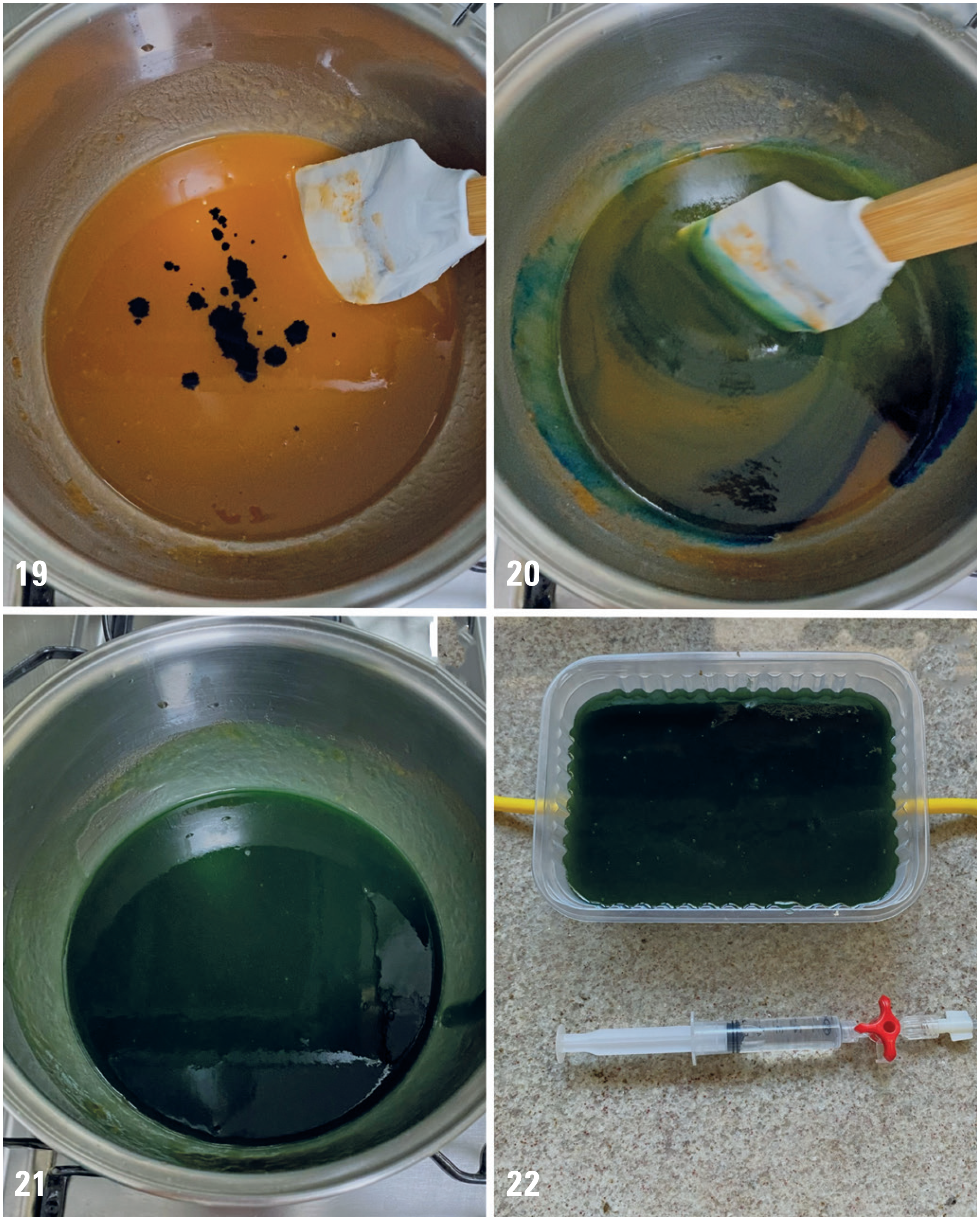
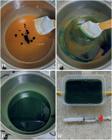
The proposed formulation resulted in a model suitable for practical use, lightweight, and without strict storage requirements. It weighs 530g, enabling transport without refrigeration. The ingredient ratio consists of 500mL of water, 60 g of gelatin, 100mL of glycerin, and 36g of psyllium, along with 10 drops of food coloring, with a total preparation time of approximately three hours (Figures 3 to 6). Furthermore, the model can be preserved for up to seven days at room temperature or up to 60 days under refrigeration.
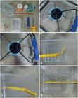
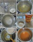
A disposable plastic container was prepared to accommodate the phantom. Using a heated corkscrew or another pointed metal object, two small lateral openings were created approximately 1.5cm from the bottom of the container. The closed end of a tubular balloon was inserted through one opening using forceps, and the procedure was repeated on the opposite side to allow the balloon to traverse the container. Care was taken to maintain the smallest possible opening to prevent leakage of the mixture.
The open end of the tubular balloon was then cut and connected to a 20cm extension set. The threaded portion of the extension set was positioned over the balloon and gently tightened to secure the connection while ensuring that the balloon material remained wrapped around the connector. The balloon was stretched inside the container, and a syringe was attached to the extension set to remove air and create a vacuum. The balloon was then slowly filled with fluid until it assumed a tubular configuration, simulating a vessel.
For preparation of the phantom matrix, 60g of unflavored gelatin was first dissolved in 100mL of cold water. The mixture was heated over medium heat, and an additional 400mL of water was gradually added while stirring until the gelatin dissolved completely. Subsequently, 100mL of glycerin was incorporated, and the mixture was maintained over medium heat until boiling. After boiling, the solution was allowed to rest for approximately 10 minutes. Psyllium (36g; Metamucil) was then added and stirred rapidly until completely dissolved. Food coloring (10 drops) was added and mixed until a homogeneous color was obtained.
The prepared mixture was poured into the container containing the balloon. The model was placed in the freezer for approximately 10 minutes to accelerate initial solidification and was then transferred to refrigeration. The phantom became ready for use after approximately 3-4 hours and could be used to simulate both in-plane and out-of-plane ultrasound-guided puncture techniques. During training, puncture practice could initially be performed using only the needle to develop eye–hand–screen coordination. The syringe could subsequently be incorporated to enhance procedural simulation. When pressure within the balloon decreased after repeated punctures, the balloon could be refilled through the extension set to allow continued use of the model.
Phase 2 – Expert panel
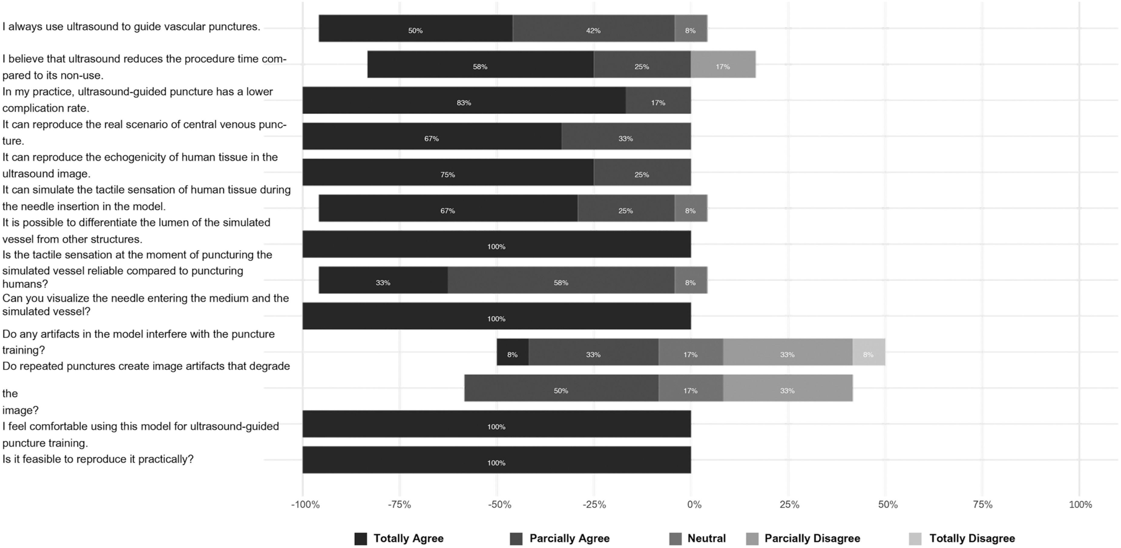
The expert panel comprised 12 specialists, including six surgeons and six non-surgeons, of whom five reported performing ultrasound-guided vascular punctures one to five times per week. The median number of punctures performed was 17.5, ranging from 5 to 100. Responses regarding the model were recorded using a Likert scale and are presented in figure 7. According to Gwet's AC2 coefficient, the agreement index among specialists was 95.5%.
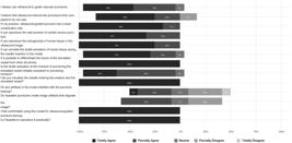
The evaluation criteria included scenario realism, echogenicity, tactile sensation, identification of the vessel lumen, image artifacts, reproducibility, needle visualization, and suitability for training. Overall, 85.83% of the evaluations were reported as positive.
DISCUSSION
In clinical practice, ultrasound-guided puncture has consistently reduced mechanical complications associated with CVA.(32,33) Simulation-based training has therefore become an important strategy to enhance patient safety. In response to this need, the present study developed a phantom with a favorable cost–benefit ratio for large-scale training, based on testing of materials previously reported in the literature. Furthermore, with the increasing use of ultrasound in medical practice, an easily reproducible teaching tool may also be incorporated into the academic setting to facilitate large-scale training of medical students, as already demonstrated by other authors.(23)
Gelatin-based models have been described as a strategy to reduce production costs,(21) which guided the selection of gelatin as the base material for the phantom developed in this study. Despite favorable results reported previously,(29,30) these models did not demonstrate ultrasound characteristics closely resembling human tissue. To improve the echographic quality of homemade models, other studies have tested materials such as psyllium(23) and cornstarch.(31) In the experiments conducted in this study, psyllium produced a satisfactory ultrasound image,(23,34) consistent with previous reports. In contrast, the cornstarch-based model(31) demonstrated acceptable ultrasound performance for puncture but showed inferior results regarding post-puncture artifacts compared with psyllium (Table 4).
Owing to difficulty in sourcing ballistic gelatin from American models,(29) a mixture of gelatin and glycerin was tested as an alternative. The addition of glycerin improved storage durability and increased resistance to biological proliferation, thereby extending refrigeration time compared with gelatin-only models.(35) Previous studies(36) have also identified agar as a suitable material for phantom production. When tested in combination with the other ingredients, agar produced satisfactory results; however, image quality deteriorated after several days of storage.
Using the final proportions, a phantom that fulfilled the initial objectives was successfully developed. In terms of cost, the model was approximately ten times less expensive than currently available commercial phantoms and comparable in price to other gelatin-based models, such as the one developed at Thomas Jefferson University (Philadelphia, PA, USA).
Regarding durability, the phantom withstood up to 100 consecutive punctures during testing, supporting its use for training. Because simulation fidelity depends on the scenario created, the proposed model supports medium-complexity training. Validation by specialists showed that 85.83% of evaluations were favorable across the assessed characteristics. The main drawback is progressive deterioration, particularly when the model is used by inexperienced trainees or stored for prolonged periods outside refrigeration.
The primary limitation of this study relates to the expert panel, which evaluated the model based on the perspective of experienced physicians rather than the intended target population. Consequently, the model may not fully reflect its use by inexperienced operators. In addition, participant selection was based on convenience sampling, which may have introduced bias into the results. Although the checklist completed by the panel was based on the literature, it has not been formally validated. Therefore, further studies are required to assess the effectiveness of the model.
CONCLUSION
The model proved to be a valuable option for simulating ultrasound-guided punctures owing to low cost, ease of transport, and the ability to reproduce realistic puncture scenarios. Further research is recommended to support large-scale training and comparative evaluations with commercial models.
DATA AVAILABILITY
The underlying data are contained within the manuscript.
REFERENCES
- 1 Crenshaw NA, Briones P, Gonzalez JM, Ortega J. A Review of Central Venous Access Using Ultrasound Guidance Technology. Adv Emerg Nurs J. 2020;42(2):119-27.
- 2 De Siqueira Simão AL, Martins Alencar G, Alcântara Garzin AC. Segurança do paciente na prática simulada durante a graduação na área da saúde. Rev Nurs Bras. 2022;25(284):6937-52.
- 3 Park O, Jeon M, Kim M, Kim B, Jeong H. The effects of a simulation-based patient safety education program on compliance with patient safety, perception of patient safety culture, and educational satisfaction of operating room nurses. Healthcare (Basel). 2023;11(21):2824.
- 4 Kumar A, Chuan A. Ultrasound guided vascular access: efficacy and safety [Internet]. Best Pract Res Clin Anaesthesiol. 2009;23(3):299-311.
- 5 Brass P, Hellmich M, Kolodziej L, Schick G, Smith AF. Ultrasound guidance versus anatomical landmarks for internal jugular vein catheterization. Cochrane Database Syst Rev. 2015;1(1):CD006962.
- 6 Shojania KG, Duncan BW, McDonald KM, Wachter RM, Markowitz AJ. Making health care safer: a critical analysis of patient safety practices. Evid Rep Technol Assess (Summ). 2001;(43):i-x, 1-668.
- 7 National Institute for Clinical Excellence. Guidance on the use of ultrasound locating devices for placing central venous catheters. [Technology appraisal guidance; no. 49]. London : National Institute for Clinical Excellence; 2002.
- 8 Bodenham A, Babu S, Bennett J, Binks R, Fee P, Fox B, et al. Association of Anaesthetists of Great Britain and Ireland: Safe vascular access 2016. Anaesthesia. 2016;71(5):573-85.
- 9 Frykholm P, Pikwer A, Hammarskjöld F, Larsson AT, Lindgren S, Lindwall R, Taxbro K, Oberg F, Acosta S, Akeson J. Clinical guidelines on central venous catheterisation. Swedish Society of Anaesthesiology and Intensive Care Medicine. Acta Anaesthesiol Scand. 2014;58(5):508-24.
- 10 Lamperti M, Bodenham AR, Pittiruti M, Blaivas M, Augoustides JG, Elbarbary M, et al. International evidence-based recommendations on ultrasound-guided vascular access. Intensive Care Med. 2012;38(7):1105-17.
- 11 Frankel HL, Kirkpatrick AW, Elbarbary M, Blaivas M, Desai H, Evans D, et al. Guidelines for the Appropriate Use of Bedside General and Cardiac Ultrasonography in the Evaluation of Critically Ill Patients-Part I: general Ultrasonography. Crit Care Med. 2015;43(11):2479-502.
- 12 Jenssen C, Brkljacic B, Hocke M, Ignee A, Piscaglia F, Radzina M, et al. EFSUMB Guidelines on Interventional Ultrasound (INVUS), Part VI - Ultrasound-Guided Vascular Interventions. Ultraschall Med. 2016;37(5):473-6.
- 13 Timsit JF, Baleine J, Bernard L, Calvino-Gunther S, Darmon M, Dellamonica J, et al. Expert consensus-based clinical practice guidelines management of intravascular catheters in the intensive care unit [Internet]. Ann Intensive Care. 2020;10(1):118.
- 14 Shime N, Hosokawa K, MacLaren G. Ultrasound Imaging Reduces Failure Rates of Percutaneous Central Venous Catheterization in Children. Pediatr Crit Care Med. 2015;16(8):718-25.
- 15 Miller JD. Editorial. Personal Disord. 2022;13(1):1-2.
- 16 Kurashima Y, Hirano S. Systematic review of the implementation of simulation training in surgical residency curriculum. Surg Today. 2017;47(7):777-82.
- 17 Tekgul ZT, Yesilnacar C, Bilgin MU, Ozkarakas H. Effectiveness of Using a Gelatine-Based Model in Ultrasonography-guided Jugular Central venous Catheter Placement Training: A Randomised Clinical Trial. J Coll Physicians Surg Pak. 2022;32(5):596-601.
- 18 Collings DG, Scullion H. Resourcing international assignees. In: International Human Resource Management. 2nd Ed. Routledge; 2008. 87-106. Pp.
- 19 Littlewood KE. High fidelity simulation as a research tool. Best Pract Res Clin Anaesthesiol. 2011;25(4):473-87.
- 20 Lerski RA, Duggan TC, Christie J. A simple tissue-like ultrasound phantom material. Br J Radiol. 1982;55(650):156-7.
- 21 García-Carpintero E, Naredo E, Vélez-Vélez E, Fuensalida G, Ortiz-Miluy G, Gómez-Moreno C. Phantoms for ultrasound-guided vascular access cannulation training: a narrative review. Med Ultrason. 2023;25(2):201-7.
- 22 Hocking G, Hebard S, Mitchell CH. A review of the benefits and pitfalls of phantoms in ultrasound-guided regional anesthesia. Reg Anesth Pain Med. 2011;36(2):162-70.
- 23 Sevak S, Lurvey B, Woodfin AA, Hothem Z, Callahan RE, Robbins J, et al. Solid, Cystic, and Tubular: Novice Ultrasound Skills Training Using a Versatile, Affordable Practice Model. J Surg Educ. 2018;75(5):1403-9.
- 24 Leenders B, Bruijnen S, Elhof J-WE. Ultrasound-guided central line placement: is a gelatine phantom a good and affordable alternative? J Surg Simul. 2017;6000:35-40.
- 25 Gwet KL. Computing inter-rater reliability and its variance in the presence of high agreement. Br J Math Stat Psychol. 2008;61(Pt 1):29-48.
- 26 Gwet KL. Handbook of Inter-Rater Reliability. The Definitive Guide to Measuring the Extent of Agreement Among Raters. Analysis of Categorical Ratings (Handbook of Inter-Rater Reliability. vol 1. Gaithersburg, MD: STATAXIS Publishing Company; 2021. 411p.
- 27 Gwet KL. On Krippendorff's Alpha Coefficient. Gaithersburg; 2015.
- 28 Amini R, Kartchner JZ, Stolz LA, Biffar D, Hamilton AJ, Adhikari S. A novel and inexpensive ballistic gel phantom for ultrasound training. World J Emerg Med. 2015;6(3):225-8.
- 29 Morrow DS, Broder J. Cost-effective, reusable, leak-resistant ultrasound-guided vascular access trainer. J Emerg Med. 2015;49(3):313-7
- 30 Selame LA, Risler Z, Zakaria SJ, Hughes LP, Lewiss RE, Kehm K, et al. A comparison of homemade vascular access ultrasound phantom models for peripheral intravenous catheter insertion. J Vasc Access. 2021;22(6):891-7.
- 31 Cheruparambath V, Sampath S, Deshikar LN, Ismail HM, Bhuvana K. A low-cost reusable phantom for ultrasound-guided subclavian vein cannulation. Indian J Crit Care Med. 2012;16(3):163-5.
- 32 Adrian M, Borgquist O, Bentzer P, Åkeson J, Spångfors M, Wrigstad J, et al. Research protocol for mechanical complications after central venous catheterisation: a prospective controlled multicentre observational study to determine incidence and risk factors of mechanical complications within 24 hours after cannulation. BMJ Open. 2019;9(10):e029301.
- 33 Smit JM, Raadsen R, Blans MJ, Petjak M, Van de Ven PM, Tuinman PR. Bedside ultrasound to detect central venous catheter misplacement and associated iatrogenic complications: a systematic review and meta-analysis. Crit Care. 2018 Mar;22(1):65.
- 34 Qurash MT, Yaacob NY, Azuan N, Khaleel YS, Zakaria R. Special ultrasound phantom for interventional training: Construction, advantages, and application. J Med Ultrasound. 2018;26(4):210-4.
- 35 Kocharyan H, Kallini J, Aida SK, Harvill M. A cost-effective alternative formulation of ultrasound phantom for vascular access instruction: cost-effective hands-on procedural training. J Vasc Access. 2018;19(3):328-9.
- 36 Earle M, Portu G, DeVos E. Agar ultrasound phantoms for low-cost training without refrigeration. Afr J Emerg Med. 2016;6(1):18-23.
Edited by
-
Associate Editor:
Ricardo Sales dos Santos Hospital Israelita Albert Einstein, São Paulo, SP, Brazil ORCID: https://orcid.org/0000-0002-7972-0355