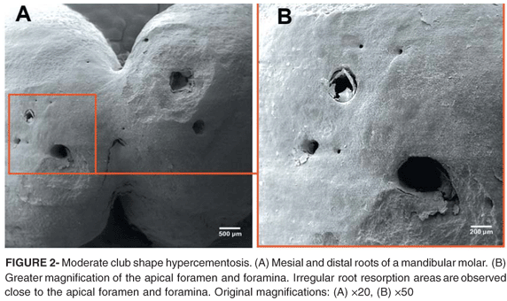
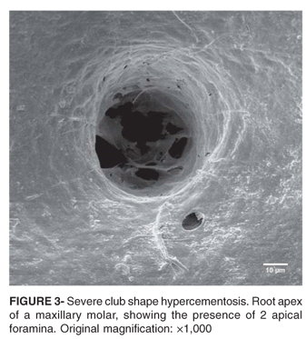
The purpose of this study was to evaluate morphological characteristics of teeth with hypercementosis that are relevant to endodontic practice. Twenty-eight extracted teeth with hypercementosis had their root apexes analyzed by scanning electron microscopy (SEM). The teeth were divided according to tooth groups and type of hypercementosis. The following aspects were examined under SEM: the contour and regularity of the root surface; presence of resorption; presence and number of apical foramina, and the diameter of the main foramen. The progression of club shape hypercementosis was directly associated with the presence of foramina and apical foramen obstruction. Cases of focal hypercementosis presented foramina on the surface, even when sidelong located in the root. Circular cementum hyperplasia form was present in 2 out of 3 residual roots, which was the highest proportion among the tooth types. The detection of a large number of foramina in the apical third of teeth with hypercementosis or even the possible existence of apical foramen obliteration contributes to understand the difficulties faced during endodontic treatment of these cases.
Hypercementosis; Root canal; Morphology; Endodontics