Abstracts
OBJECTIVE: The aim of this study was to determine the differences between the skeletal ages estimated by TW2 and TW3 methods through their RUS and Carpal systems. MATERIAL AND METHODS: A sample of two hundred and forty hand and wrist radiographs of male and female Brazilian children aged 84-199 months was evaluated by five observers. The Dunnet test was performed for statistical analysis. RESULTS: Results showed higher skeletal ages estimated by TW2RUS than TW3RUS and Carpal for both genders. For girls a statistically significant difference (p<0.05) was observed between TW2RUS and TW3RUS over the entire age range. For boys this difference was observed from 108 months onwards. In general RUS skeletal ages were higher than the chronological age and Carpal skeletal ages for both genders. The overestimation of chronological age was smaller for TW3RUS than for TW2RUS, and this last system showed a statistically significant difference regarding chronological age over the entire age range for girls, whereas for boys this difference was seen from 132 months onwards. For girls TW3 RUS and Carpal showed a significant difference regarding chronological age in the oldest age groups; in boys TW3RUS did not show a significant difference regarding chronological age. For Carpal, these results were more variable. CONCLUSION: It seems reasonable to recommend the use of the TW3 system for the studied Brazilian population.
Skeletal maturity; Skeletal age; Hand and wrist radiograph
OBJETIVO: O objetivo deste estudo foi determinar as diferenças entre as idades esqueletais estimadas pelos métodos TW2 e TW3, usando os sistemas RUS e Carpal. MATERIAL E MÉTODOS: Uma amostra de 240 radiografias de mão e punho de crianças brasileiras de ambos os sexos com idade cronológica entre 84 e 199 meses foram avaliadas por cinco observadores. Para análise estatística dos dados foi aplicado o Teste de Dunnet. RESULTADOS: Os resultados demonstraram que as idades esqueletais estimadas pelo método TW2RUS foram mais avançadas que aquelas estimadas pelos métodos TW3RUS e Carpal, para ambos os sexos. Para o sexo feminino, uma diferença estatisticamente significante (p<0,05) foi observada entre os métodos TW2RUS e TW3RUS em todas as faixas etárias estudadas. No sexo masculino, essa diferença foi observada a partir de 108 meses em diante. Em geral, idades esqueletais estimadas pelo método RUS são maiores que a idade cronológica e também maiores que a idade esqueletal estimada pelo método Carpal em ambos os sexos. A superestimativa da idade cronológica é menor quando se aplica o método TW3RUS do que ao se utilizar o método TW2RUS, e esse último sistema mostrou uma diferença estatisticamente significante em relação a idade cronológica em todas as faixas etárias das meninas, enquanto para os meninos isso foi observado apenas a partir dos 132 meses em diante. No sexo feminino, houve uma diferença significante em relação a idade cronológica no grupo mais velho quando se comparou TW3RUS e Carpal; o que não foi observado no sexo masculino. No método Carpal os resultados foram mais variados. CONCLUSÃO: Diante dos resultados, os autores consideraram mais razoável a utilização do método TW3 para a estimativa da idade esqueletal em pacientes brasileiros.
Maturação esquelética; Idade óssea; Radiografia de mão e punho
ORIGINAL ARTICLES
Comparison of TW2 and TW3 skeletal age differences in a Brazilian population
Estudo comparativo das idades ósseas estimadas pelos métodos TW2 e TW3 numa população brasileira
Ana Isabel OrtegaI; Francisco Haiter-NetoII; Gláucia Maria Bovi AmbrosanoIII; Frab Norberto BóscoloII; Solange Maria AlmeidaII; Marcia Spinelli CasanovaIV
IAssistant Professor. Department of Oral Radiology, Dental School, Universidad del Zulia, Maracaibo, State of Zulia, Venezuela
IIAssociate Professor. Department of Oral Radiology, Piracicaba Dental School, State University of Campinas, São Paulo, Brazil
IIIAssociate Professor. Department of Social Dentistry, Piracicaba Dental School, State University of Campinas, São Paulo, Brazil
IVDDS, MSc. Department of Oral Radiology, Piracicaba Dental School, State University of Campinas, São Paulo, Brazil
Corresponding address Corresponding address: Prof. Dr. Francisco Haiter Neto Faculdade de Odontologia de Piracicaba UNICAMP - Área de Radiologia Odontológica Av. Limeira, 901 - Areião - 13414-901. Piracicaba, SP Telefone/Fax: (19) 3412 5327 E-mail: haiter@fop.unicamp.br
ABSTRACT
OBJECTIVE: The aim of this study was to determine the differences between the skeletal ages estimated by TW2 and TW3 methods through their RUS and Carpal systems.
MATERIAL AND METHODS: A sample of two hundred and forty hand and wrist radiographs of male and female Brazilian children aged 84-199 months was evaluated by five observers. The Dunnet test was performed for statistical analysis.
RESULTS: Results showed higher skeletal ages estimated by TW2RUS than TW3RUS and Carpal for both genders. For girls a statistically significant difference (p<0.05) was observed between TW2RUS and TW3RUS over the entire age range. For boys this difference was observed from 108 months onwards. In general RUS skeletal ages were higher than the chronological age and Carpal skeletal ages for both genders. The overestimation of chronological age was smaller for TW3RUS than for TW2RUS, and this last system showed a statistically significant difference regarding chronological age over the entire age range for girls, whereas for boys this difference was seen from 132 months onwards. For girls TW3 RUS and Carpal showed a significant difference regarding chronological age in the oldest age groups; in boys TW3RUS did not show a significant difference regarding chronological age. For Carpal, these results were more variable.
CONCLUSION: It seems reasonable to recommend the use of the TW3 system for the studied Brazilian population.
Uniterms: Skeletal maturity; Skeletal age; Hand and wrist radiograph.
RESUMO
OBJETIVO: O objetivo deste estudo foi determinar as diferenças entre as idades esqueletais estimadas pelos métodos TW2 e TW3, usando os sistemas RUS e Carpal.
MATERIAL E MÉTODOS: Uma amostra de 240 radiografias de mão e punho de crianças brasileiras de ambos os sexos com idade cronológica entre 84 e 199 meses foram avaliadas por cinco observadores. Para análise estatística dos dados foi aplicado o Teste de Dunnet.
RESULTADOS: Os resultados demonstraram que as idades esqueletais estimadas pelo método TW2RUS foram mais avançadas que aquelas estimadas pelos métodos TW3RUS e Carpal, para ambos os sexos. Para o sexo feminino, uma diferença estatisticamente significante (p<0,05) foi observada entre os métodos TW2RUS e TW3RUS em todas as faixas etárias estudadas. No sexo masculino, essa diferença foi observada a partir de 108 meses em diante. Em geral, idades esqueletais estimadas pelo método RUS são maiores que a idade cronológica e também maiores que a idade esqueletal estimada pelo método Carpal em ambos os sexos. A superestimativa da idade cronológica é menor quando se aplica o método TW3RUS do que ao se utilizar o método TW2RUS, e esse último sistema mostrou uma diferença estatisticamente significante em relação a idade cronológica em todas as faixas etárias das meninas, enquanto para os meninos isso foi observado apenas a partir dos 132 meses em diante. No sexo feminino, houve uma diferença significante em relação a idade cronológica no grupo mais velho quando se comparou TW3RUS e Carpal; o que não foi observado no sexo masculino. No método Carpal os resultados foram mais variados.
CONCLUSÃO: Diante dos resultados, os autores consideraram mais razoável a utilização do método TW3 para a estimativa da idade esqueletal em pacientes brasileiros.
Unitermos: Maturação esquelética; Idade óssea; Radiografia de mão e punho.
INTRODUCTION
The Tanner-Whitehouse method (TW) assesses specific ossification centers of the hand and wrist through two systems, RUS (radio, ulna and selected metacarpals and phalanges) and Carpal, which analyzes the carpal bones except for the pisiform. The individual bones are matched to a series of written criteria describing eight or nine standard stages, labeled A to H or I, through which each bone passes in its progression to maturity. Each stage is defined by up to three criteria and is assigned a specific score. The sum of these scores results in a skeletal maturity score (SMS) that can be transformed into skeletal age1.
In order to improve accuracy and reproducibility of the TW method, the scoring system, maturity stages, skeletal ages and even the equations for the prediction of adult height have been modified throughout the years. This process led to the publication of the new version of this method, termed TW3.
This system maintains the description and manual rating of the bones' stages, thus the RUS and Carpal scores are exactly the same in TW3 as in TW2. Also, the 20-bone score was abolished and the reference values and charts for RUS were changed and based on data from North American children9.
Considering these changes, this study compares the differences in Tanner-Whitehouse skeletal ages as derived from TW2 and TW3 systems, in Brazilian children.
MATERIAL AND METHODS
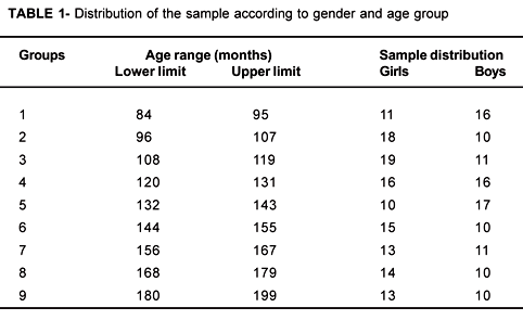
Two hundred and forty hand and wrist radiographs from Brazilian children (111 boys and 129 girls) aged 84-199 months were selected from the archives of the Department of Radiology (Piracicaba Dental School). Radiographs were divided into nine groups based on chronological age for each gender, with an interval of 12 months between them (Table 1).
Five observers assessed independently each radiograph without knowledge of the chronological age of the child, according to the TW3 method (RUS and Carpal). RUS skeletal ages were calculated from two different sources: TW2 standards10 (TW2RUS) and TW3 standards9 (TW3RUS). For the Carpal system, skeletal ages were calculated using TW3.
The Dunnet test was performed in order to determine if there was a statistically significant difference between the skeletal ages estimated by all methods and the chronological age. Furthermore an F-test was performed in order to look for an actual statistical difference between TW2RUS and TW3RUS.
RESULTS
Tables 2 and 3 show the means and the standard deviations of TW2RUS, TW3RUS and Carpal skeletal ages as well as the chronological ages of both genders.
For girls, these results are shown in Table 2. There was a statistically significant difference between TW2RUS and TW3RUS mean skeletal ages over the entire age range. A statistically significant difference between TW2RUS skeletal ages and chronological age was also observed. TW3RUS showed a statistical difference regarding the chronological age of the sixth age group onwards except for the eighth group. For the Carpal, there was a statistically significant difference regarding the chronological age from group seven to group nine.
For boys (Table 3) there was a statistically significant difference between TW2RUS and TW3RUS from the third age group to the last one. Regarding chronological age, there was a statistical difference between TW2RUS skeletal ages and chronological age from group five to group nine. Carpal showed a significant difference regarding chronological age in age groups one, two, five and nine.
Table 4 shows the mean differences between TW2RUS minus TW3RUS, TW2RUS minus Carpal and TW3RUS minus Carpal for both genders.
Positive means in favor of TW2RUS bone ages were observed for both genders, except for groups one and two for boys when calculated by TW2RUS minus TW3RUS. For TW3RUS minus Carpal, negative means in favor of Carpal are observed in groups one, two and three for girls and in groups four, five and six for boys.
Figures 1 and 2 show the relationship between TW2, TW3 and Carpal skeletal ages and the chronological age.
For girls (Figure 1) TW2RUS and TW3RUS overestimated chronological age, this overestimation was greater for TW2RUS than for TW3RUS. Carpal score underestimated chronological age; its skeletal ages were smaller than TW2RUS over the entire age range and were higher than TW3RUS from 84 to 119 months.
DISCUSSION
Some authors have compared TW1 and TW2 versions in populations of different origin 3,5,11 and verified that the skeletal ages estimated by the TW1 system were, in general, higher than TW2 skeletal ages, probably because in the TW2 system the adult score is reached one year earlier than in TW1. This study compares the TW2 system, which is based on the original British population of the TW method, and the TW3 system that presents a new standardized RUS score using a North American sample. The correspondence of the skeletal age estimated by all systems with the chronological age of the studied individuals was also determined.
In this research, the authors observed that the female skeletal ages estimated by TW2RUS were higher than the chronological age, as reported by Lejarraga, et al.4 The skeletal ages estimated by TW3RUS and Carpal were smaller than TW2RUS skeletal ages, but they were similar to the chronological age (Table 2, Figure 1). The standard deviation for TW2RUS and TW3RUS mean skeletal ages observed in the ninth age group reflects that most girls reached the adult stage in this group. Therefore, there was a minimum variation of the estimated skeletal ages with regard to the mean skeletal age.
For boys the skeletal ages estimated by TW2RUS were higher than the chronological age from the fifth age group onwards. Whereas TW3RUS and Carpal skeletal ages were closer than TW2RUS to the chronological age over the entire age range (Table 3, Figure 2).
Among genders, girls showed higher skeletal ages than boys for all systems, indicating that there is an actual precocity of females regarding skeletal maturation. 2,6,7,8 Molinari, et al.6 reported in 2004 that girls reach the TW3 final score about 2 years earlier than boys, and the skeletal maturation is more rapid in girls than in boys soon in childhood.
According to table 4, the differences between TW2RUS and TW3RUS were in average 11 months for girls, as related by Tanner, et al.9. For boys the differences from -0.17 to 8.04 months were observed in the youngest age groups, and a mean difference of 13.4 months from the fifth age group onwards. These differences between TW2RUS and TW3RUS are possibly due to the environmental background of the populations used for the development of the methods, as well as to the inherent characteristics of the studied individuals. It seems that the maturation pattern observed in the studied children is more alike the North American standards, meaning that Brazilian children are ahead of the British children regarding their skeletal maturation.
As table 4 shows, for girls the difference between the mean skeletal age assessed by TW2RUS and Carpal assumed higher values from the fifth group (aged 132-143 months) onwards than from the fourth group (aged 120-131 months) downwards; this probably indicates that the majority of girls were rated as adults by both systems from this age on. The adult score is reached at 16 years and 13 years by the TW2RUS system and by the Carpal system, respectively; this fact can explain the large discrepancy between the means of these two systems. For boys, the difference between the mean skeletal ages assessed by TW2RUS and Carpal assumed higher values from the seventh group (aged 156-167 months) onwards than from the sixth group (aged 144-155 months) downwards. However, the differences between the TW2RUS and the Carpal for boys were not so marked as those seen in girls, probably because the majority of boys did not reach the adult stage at the age range in the study.
Between TW3 and Carpal, the differences were smaller than the differences found between TW2RUS and Carpal for both genders. Nevertheless, since Carpal, in general, estimated smaller skeletal ages than TW2RUS and TW3RUS and the chronological age, it seems reasonable to state that there is an actual delay in the development of carpal bones compared to metacarpal and phalanges.
CONCLUSIONS
There was a statistically significant difference between TW2RUS and TW3RUS mean skeletal ages for both genders; however, for boys it was only observed from the third age group on.
The skeletal ages estimated by the TW2RUS method were higher than the TW3RUS method for both genders, except for the first two groups for boys.
Comparison to TW2RUS method, the skeletal ages estimated by TW3RUS method were closer to Brazilian chronological ages; it seems reasonable to recommend the use of the TW3 system in Brazilian population.
Received: July 05, 2005
Modification: October 07, 2005
Accepted: December 15, 2005
- 1- Gilli G. The assessment of skeletal maturation. Horm Res. 1996;45:49-52.
- 2- Guzzi BSS, Carvalho LS. Estudo da maturação óssea em pacientes jovens de ambos os sexos através de radiografias de mão e punho. Ortodontia. 2000;33:49-58.
- 3- Kimura K. Skeletal maturity of the hand and wrist in Japanese children by the TW2 method. Am J Phys Anthropol. 1971;4:353-6.
- 4- Lejarraga H, Guimarey L, Orazi V. Skeletal maturity of the hand and wrist of healthy Argentinean children aged 4-12 years, assessed by the TW II method. Ann Hum Biol. 1997;24:257-61.
- 5- Malina RM, Little BB. Comparison of TW1 and TW2 skeletal age differences in American black and in Mexican children of 6-13 years of age. Ann Hum Biol. 1981;8:543-8.
- 6- Molinari L, Gasser T, Largo RH. TW3 Bone age: RUS/CB and gender differences of percentiles for score and score increments. Ann Hum Biol. 2004;31:421-35.
- 7- Pryor JW. Differences in the time of development of centers of ossification in the male and female skeleton. Anat Rec. 1923;25:257-73.
- 8- Roche AF, Rohmann CG, French NY, Davila GH. Effect of training on replicability of assessments of skeletal maturity (Greulich-Pyle). AJR Am J Roentgenol. 1970;108:511-5.
- 9- Tanner JM, Whitehouse RH, Cameron N, Marshall WA, Healy MJR, Goldstein NH. Assessment of skeletal maturity and prediction of adult height (TW3 method). 3rd ed. London: W.B Saunders; 2001.
- 10- Tanner JM, Whitehouse RH, Marshall WA, Healy MJR, Goldstein H. Assessment of skeletal maturity and prediction of adult height (TW2 method). 2 ed. London: Academic Press;1983:108p.
- 11- Van Venrooij-Ysselmuiden ME, Van Ipenburg A. Mixed longitudinal data on skeletal age from a group of Dutch children living in Ultrech and surroundings. Ann Hum Biol. 1978;5:359-80.
Publication Dates
-
Publication in this collection
08 June 2006 -
Date of issue
Apr 2006
History
-
Accepted
15 Dec 2005 -
Received
05 July 2005 -
Reviewed
07 Oct 2005


