Abstracts
Two quite dyspneic HIV positive patients were admitted to the Emergency Room; they presented clinical signs and images suggesting pericardial effusion. The analysis of an initial liquid puncture did not show any specificity and the patients did not exhibit any clinical improvement. Both patients were submitted to a subxiphoid pericardial window, all the effusion liquid was drained, and a biopsy of the pericardium tissue was completed, revealing a granulomatous process. Immediately after the onset of specific treatment, the patients showed a good evolution. Such findings draw attention to a high possibility of pericardial suffusion in AIDS patients being tuberculosis, particular if one considers the high prevalence of this disease in Brazil. The results also showed that the opening of a subxiphoid pericardial window and the specific triple scheme was a procedure that led to good therapeutic evolution in these patients.
Pericarditis; AIDS; Tuberculosis; Pericardial window
Dois pacientes portadores do vírus HIV deram entrada no serviço de emergência bastante dispnéicos, exibindo sinais clínicos e de imagens sugestivos de derrame pericárdico. Realizada inicialmente a punção do líquido, sua análise não mostrou especificidade e os doentes não apresentavam evidência de melhora clínica. Foram, então, submetidos a uma janela pericárdica subxifóidea, foi drenado todo o líquido de efusão e realizada a biópsia do tecido pericárdico, o que revelou processo granulomatoso. Logo após o início do tratamento específico, os pacientes apresentaram boa evolução. Tais achados chamaram a atenção para a etiologia tuberculosa como causa de sufusão pericárdica em portadores da síndrome de imunodeficiência adquirida. Essa associação pode ser mais importante no Brasil, onde existe alta prevalência de tuberculose. Os resultados mostraram também que a realização de uma janela pericárdica subxifóidea permitiu boa drenagem do fluido e, junto com a ministração do esquema tríplice, possibilitou a boa evolução dos pacientes.
Pericardite; AIDS; Tuberculose; Janela pericárdica
CASE REPORT
Tuberculous pericarditis in acquired immune deficiency syndrome patients* * Study carried out at Hospital Ipiranga, São Paulo, SP.
Pericardite tuberculosa em portadores da síndrome de imunodeficiência adquirida
Ruggero Bernardo GuidugliI; Paul Albert HamrickII; Nancy Figueiroa de RezendeII
ISurgeon of the Thoracic Surgery Service. Master in Surgical Technique and Experimental Surgery
IIInfectologist
Correspondence Correspondence to Ruggero Bernardo Guidugli Amiamspe, Rua Borges Lagoa, 1.855 04038-034 São Paulo, SP
ABSTRACT
Two quite dyspneic HIV positive patients were admitted to the Emergency Room; they presented clinical signs and images suggesting pericardial effusion. The analysis of an initial liquid puncture did not show any specificity and the patients did not exhibit any clinical improvement. Both patients were submitted to a subxiphoid pericardial window, all the effusion liquid was drained, and a biopsy of the pericardium tissue was completed, revealing a granulomatous process. Immediately after the onset of specific treatment, the patients showed a good evolution. Such findings draw attention to a high possibility of pericardial suffusion in AIDS patients being tuberculosis, particular if one considers the high prevalence of this disease in Brazil. The results also showed that the opening of a subxiphoid pericardial window and the specific triple scheme was a procedure that led to good therapeutic evolution in these patients.
Key words: Pericarditis. AIDS. Tuberculosis. Pericardial window.
RESUMO
Dois pacientes portadores do vírus HIV deram entrada no serviço de emergência bastante dispnéicos, exibindo sinais clínicos e de imagens sugestivos de derrame pericárdico. Realizada inicialmente a punção do líquido, sua análise não mostrou especificidade e os doentes não apresentavam evidência de melhora clínica. Foram, então, submetidos a uma janela pericárdica subxifóidea, foi drenado todo o líquido de efusão e realizada a biópsia do tecido pericárdico, o que revelou processo granulomatoso. Logo após o início do tratamento específico, os pacientes apresentaram boa evolução. Tais achados chamaram a atenção para a etiologia tuberculosa como causa de sufusão pericárdica em portadores da síndrome de imunodeficiência adquirida. Essa associação pode ser mais importante no Brasil, onde existe alta prevalência de tuberculose. Os resultados mostraram também que a realização de uma janela pericárdica subxifóidea permitiu boa drenagem do fluido e, junto com a ministração do esquema tríplice, possibilitou a boa evolução dos pacientes.
Descritores: Pericardite. AIDS. Tuberculose. Janela pericárdica.
Abbreviations used in this article
AIDS Acquired immunodeficiency syndrome
HIV Human immunodeficiency virus
KB Koch's bacillus
INTRODUCTION
Currently, the large amount of pericardial effusions are related to AIDS pathologies (1,2) and, due to the high prevalence in our country, tuberculosis must always be recalled as one etiology of pericarditis in patients with HIV.
However, diagnostic difficulties may occur by the simple analysis of pericardial liquid obtained by puncture, which may not show specificity. This fact leads us to perform a biopsy.
This situation took place with two of our patients. Their reports will help us to conduct this association better.
CASE REPORTS
Case 1
31 year-old HIV-positive man, being treated with anti-viral drugs as well as trimethoprim and sulfamethoxazole for pneumocystosis. Admitted at the emergency room with intense dyspnea.
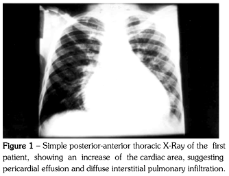
He had a fever (T 37.6º C), was tachycardiac (P 120 bpm) and the simple thoracic radiography showed an increase of the cardiac area with diffuse interstitial pulmonary infiltration, which was interpreted as pneumocystosis and treated with trimethoprim and sulfamethoxazole (Figure 1).
We hypothesized a pericardial effusion, which was confirmed by echocardiographic exam.
Having been submitted to a pericardial puncture, approximately 150 ml of a citric-yellow liquid was removed, showing 54% of lymphocytes, 40% of neutrophils, 4.2 g/l of protein, 18 g/l of glucose. Negative ADA. A sample was sent for KB culture. Due to the worsening of the dyspnea and the clinical features, a subxiphoid pericardial window and biopsy were performed under general anesthesia and approximately 280 ml of liquid was removed and a drain was kept in the mediastinum.
There was a fast improvement of the dyspnea and the result of the biopsy revealed tuberculous pericarditis (Figure 2), allowing the introduction of a triple schedule, resulting in a significant and fast improvement of the patient's status.
KB culture took 45 days and was also positive.
The patient was followed-up for 9 months and there was no relapse of the symptoms.
Case 2
45 year-old man, admitted into the emergency room with cough and intense shortness of breath. At the clinical examination he was quite dyspneic, tachycardiac and with fever (T 37.9º C). The simple thoracic radiography showed increased cardiac area and multiple sites of pulmonary condensation.
Computer assisted tomography evidenced zones of alveolar condensation and reticular-node interstitial infiltration (Figure 3), which were interpreted as probable tuberculosis, not confirmed by the bacterioscopic exam.
HIV serology was positive (Elisa and Western blot) and the pericardial effusion was confirmed by echocardiographic exam.
The patient was submitted to a pericardial puncture with removal of approximately 120 ml of a yellow-citric liquid, with 40% of neutrophils, 60% of lymphocytes, 26 g/l of glucose, 3.8 g/l of proteins, negative ADA and negative bacteriology.
Due to a progressive increase of the dyspnea, he was submitted to a subxiphoid pericardial window under general anesthesia, removing 240 ml of a yellow-citric liquid. After the surgery, there was a clear improvement of the dyspnea and the histopathological exam also revealed tuberculous pericarditis.
After the first days of triple schedule, there was a fast improvement of the patient's status, as well as of the pulmonary features. During a nine month follow-up, there was no sign of relapse of the disease.
DISCUSSION
Before AIDS appearance, pericardial effusions were mainly caused by uremia, malignancies and viral pericarditis (1).
Nowadays, in large urban centers, etiology of pericarditis is mainly related to the AIDS-associated pathologies (2). In African countries, tuberculosis is responsible for 100% of pericarditis cases in individuals with this syndrome (3,4).
We believe that, in Brazil, tuberculous pericarditis has a major role among pericardial effusions of HIV. However, due to the possibility of association with multiple opportunistic agents, which may also cause pericardial effusion and the low specificity of liquid analysis, there is still some difficulty to support the diagnosis.
Pericardial effusion may be the main sign of localized or spread tuberculosis and, usually, results in endangering the pericardial sac by mediastinal lymphonodus contaminated by mycobacterium (5,6).
Usually, pericardial liquid is a yellow-citric exudate with high number of lymphocytes and low glucose. Its culture does not always result bacillus positive. If not conveniently emptied, it will evolve to formation of fibrin, septation and granuloma with adherence and thickening of leaflets, developing to constrictive chronic pericarditis. Corticoids are known to be beneficial, hindering liquid re-accumulation, but the literature still lacks data on the prevention of constrictive pericarditis (7).
The complete and permanent drainage of the effusion liquid would be the best way to avoid future constriction of the heart chambers (8).
It is therefore essential to remove the effusion liquid and to perform the histopathologic exam of the pericardium for diagnosis confirmation.
The subxiphoid pericardial window is a low morbidity surgical procedure; it allows a total drainage of the effusion liquid and the removal of a fragment the impaired tissue. In patients with precarious conditions, it can be done under local anesthesia (9-11).
In a review of 29 AIDS cases with pericardial effusion, submitted to a pericardial window or to a thoracotomy, Flum et al. (12) concluded that pericarditis in AIDS patients is associated with a bad prognosis. Such surgical procedures would not be beneficial to the patients due to post-operative complications.
Our patients were quite dyspneic and the pericardial window allowed a fast clinical improvement. The biopsy allowed the adequate treatment, with a good evolvement of the patients.
We believe that, in our environment, in HIV patients who develop a pericardial effusion and present an inconclusive result in the liquid obtained by puncture, there is a great possibility to treat tuberculosis. In this case, the subxiphoid pericardial window constitutes an adequate conduct.
This procedure would allow a biopsy to be performed, shaping the diagnostic and the total and permanent drainage of the effusion liquid, preventing constrictive pericarditis.
Received for publication on 12/9/02
Accepted after revision on 2/12/03
- 1. Chen Y, Brennessel D, Walters J, Johnson M, Rosner F, Raza M. Immunodeficiency virus associated pericardial effusions Report of 40 cases and review of the literature. Am Heart J 1999;137:516-20.
- 2. Schiller M, Gordon AS, Eisemberg M. HIV associated pericardial effusions. Chest 1992;102:956-8.
- 3. Cegielski GJ, Ramiya K, Lallinger GJ, Mtulia IA, Mbaga IM. Pericardial disease and human immunodeficiency virus in Dar es Salaam, Tanzania. Lancet 1990;335:209-12.
- 4. Cegielski JP, Lwakatare J, Dukes CS, Lema LE, Lallinger GJ, Kitinya J, et al. Tuberculous pericarditis in Tanzania patients with HIV infection. Tuber Lung Dis 1994;775:429-34.
- 5. Lewis W. Aids: cardiac findings from 115 autopsies. Prog Cardiovasc Dis 1989;32:207-15.
- 6. Klatt EC, Meyer PR. Pathology of the heart in AIDS. Arch Lab Pathol Med 1988;112:114-6.
- 7. Strang H, Kalxaza HHS, Gibson DG. Controlled clinical trial of complete open surgical drainage and of prednisolone in treatment of tuberculous pericardial effusion in T. Lancet 1988;2:759-64.
- 8. Desai HN. Tuberculous pericarditis: a review of 100 cases. S Afr Med J 1979;5:877-80.
- 9. Millis SA, Jukian S, Hollyday RH, Visteu-Johansen J, Case LD, Hudspeth AS, et al. Subxiphoid pericardial window for pericardial effusion disease. J Cardiovasc Surg 1989;30:768-73.
- 10. Lennin BH, Aaron BL. The subxiphoid pericardial window. Surg Gynecol Obstet 1982;155:804-6.
- 11. Fontenelle LJ, Cuello L, Dooley BN. Subxiphoid pericardial window. A simple and safe method for diagnosing and treating acute and chronic pericardial effusions. J Thorac Cardiovasc Surg 1971;62:95-7.
- 12. Flum DR, Mc Ginin Jr GT, Tyras DH. The role of the "pericardial window" in AIDS. Chest 1995;107:1522-25.
Publication Dates
-
Publication in this collection
23 June 2003 -
Date of issue
Apr 2003
History
-
Accepted
02 Dec 2003 -
Received
12 Sept 2002


