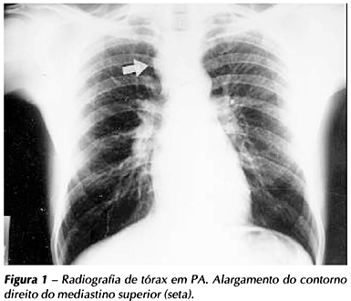
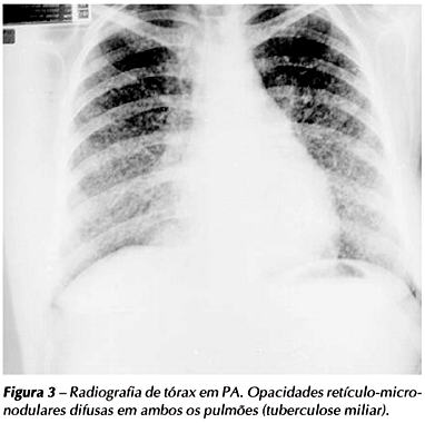
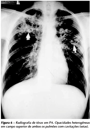
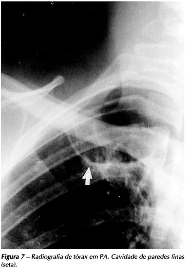
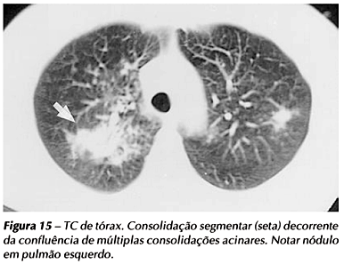
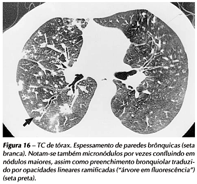
Tuberculosis is a disease of high incidence and prevalence in Brazil. Imaging methods can reveal signs suggestive of tuberculosis activity or sequelae. Chest radiographs can reveal active lung tuberculosis through consolidations, cavitations, interstitial patterns (nodular and reticulo-nodular), mediastinal or hilar lymphadenopathy and pleural effusions. Images compatible with the active disease, such as centrilobular nodules segmentarily distributed, thick-walled cavities, thickened bronchial or bronchiolar walls, bronchiectasis and lymphadenopathy can be observed by computerized tomography. Thin-walled cavities, traction bronchiectasis, parenchymal bands, emphysema and mosaic pattern are signs suggestive of inactive disease. Gallium-67 citrate scyntigraphy is a complementary method useful in the detection of infectious diseases, including tuberculosis, especially in immunocompromised patients. Inhalation / perfusion analyses are used in the pre-operative assessment of patients carrying tuberculosis sequelaes and multiresistant tuberculosis. Positron emission tomography with fluorine-18 labeled deoxyglucose allows the detection of the inflammatory process that takes place during the active stage of tuberculosis and may persist, not so intense, after specific treatment is over. Imaging methods are valuable tools to be used in the diagnosis and follow up of pulmonary tuberculosis.
Tuberculosis; Radiography; Computed tomography; Scintigraphy; Emission-computed tomography

