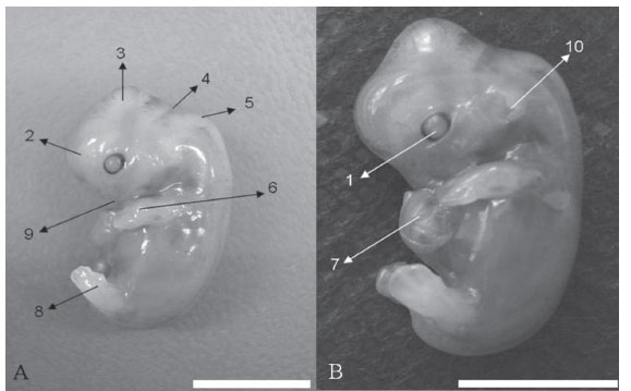
The aim of this study was to perform an embryology analysis of South-American hystricomorph rodents (paca, agouti, capybara, and rock cavy) to compare these data with the ones from other rodents and with the human embryologic morphogenesis. We used 8 samples of rodents, 2 embryos for each species, collected during early gestation. The embryos were removed from the pregnant uterus through partial ovarysalpingohysterectomy, followed by fixation in 4% paraformaldehyde solution. For crow-rump measurements, we had as reference the nuchal crest at one extremity and the last sacral vertebra on the opposite side. The embryos examined showed the following morphological characteristics: Division of the branchial arches, the cranial neuropore open in some embryos studied, the cranial curvature developed, and the somites delimited and distinct. The hindlimb and forelimb were in development, and showed "row" form; so we found the cardiac impression and the liver region. In the caudal region we observed the head-flow curve, the optic vesicle with and without pigmentation of the retina, the opening of neural tube in the fourth ventricle region of the brain, the nasal cavity, and the encephalic vesicles formation. We conclude that the embryo development of hystricomorph rodents can be compared with the morphogenesis in rats, guinea pigs, rabbits and humans in the early stages of development, being aware of the particularities for each species, in addition to the implementation of reproductive technology, especially of embryos, what requires knowledge of the pre-implantation stages for the different development phases.
Embryo development; hystricomorph rodents; South-American rodents

 South-American hystricomorphs: a comparative analysis of embryological development


