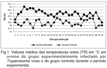
Four adult sheep (number 1, 2, 3 and 4), all males, were inoculated intravenously with 1ml of blood containing 1.25x10(5) trypomastigotes of Trypanosoma vivax, and Sheep 5, 6, 7 and 8 were used as control. After infection, clinical exams considering rectal temperature, respiratory and cardiac frequencies, and parasitaemia were recorded daily for a 30-day experiment period. Blood samples were obtained for 5-day intervals to hematocrit analysis. At the end of the experimental period, the sheep were orquiectomized. Testes and epididymides from these animals were studied anatomopathologically. Samples from these tissues of Sheep 1, 4 and 5 were taken to polymerase chain reaction (PCR). Clinical parameters remained for the infected group above the values observed in the control group during the experimental period. Parasitaemia was observed on day 3 post-infection, and the highest values occurred between day 6 and 10, and day 15 and 18 post-infection. Sheep 1 and 4 showed severe anemia on day 25 post-infection. All sheep of the infected group showed flabby and palid testes. Histologically, moderate to severe testicular degeneration, multifocal epididymitis and hyperplasia of epididymal epithelium were observed. The result of T. vivax PCR analysis in the testes and epididymal tissues was positive in 100% of the samples of the experimentally infected sheep. Epididymal and testicular lesions associated with the presence of the parasite in these tissues, shown by PCR, suggest the participation of T. vivax in the pathophysiological mechanism of reproductive damage.
Trypanosomosis; small ruminant; PCR; anatomopathology; testicular degeneration; epididymitis; extravascular foci

 Trypanosoma vivax in testicular and epidydimal tissues of experimentally infected sheep


