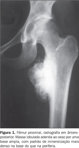
OBJECTIVE: To evaluate the most significant features of parosteal osteosarcoma and to describe the most frequent findings on conventional radiology. MATERIALS AND METHODS: A retrospective study was performed including 26 cases of patients with parosteal osteosarcoma from the archives of "Clube do Osso", Rio de Janeiro, RJ, Brazil, with analysis of main clinical and radiological findings. RESULTS: The disease was prevalent in female patients in the third decade of life. Main clinical findings were the increase in volume on the site of the tumor (77% of cases) and local pain (68% of cases). The most frequent site of tumor was the popliteal fossa (40%), and metaphyseal involvement has occurred in 92% of cases. The most frequent radiological findings were densely mineralized lesions on juxtacortical locations, and irregularly thickened adjacent host cortex (92.3%), with adherence areas being observed in 88.5% of cases, besides lobular (50%) or irregular (38.5%) tumor margins. Also, a radiolucent line between the tumor and the adjacent bone (48%), a denser mineralization on the basis than in the periphery of the tumor (42.3%), and a small rate of periosteal reaction (15.4%) were found. CONCLUSION: Although computed tomography and magnetic resonance imaging are important modalities for identifying some aspects of parosteal osteosarcoma, conventional x-ray is essential in the initial evaluation of this type of lesion, most frequently allowing differential diagnosis with other surface bone lesions.
Parosteal osteosarcoma; Bone radiology


