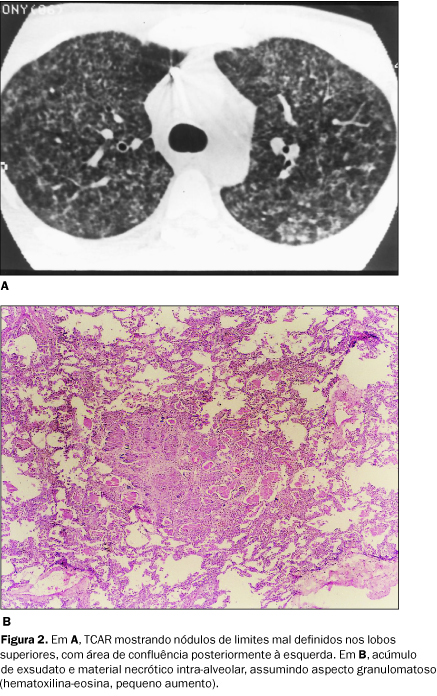
We present the main findings observed on the high-resolution computed tomography examinations of 15 patients with acquired immunodeficiency syndrome and Pneumocystis carinii pneumonia. The high-resolution computed tomography and autopsy findings of 5 patients were also compared. The most frequently observed high-resolution computed tomography patterns were ground-glass attenuation, consolidation areas, crazy-paving pattern and cysts. Nodules and intralobular reticulation were less frequently observed. Ground-glass attenuation and consolidation areas corresponded to alveolar filling with inflammatory exsudate. Thickening of the interlobular septa was due to cell infiltration and edema. One patient presented interlobular reticulation, and the pathology study revealed alveolar septa thickening due to cell infiltration and fibrosis. Nodules observed in one of the patients corresponded to a patchy intraalveolar accumulation of microorganisms and inflammatory cells forming a "granulomatous" pattern.
Pneumocystis carinii pneumonia; Acquired immunodeficiency syndrome; High-resolution computed tomography; Anatomopathology


