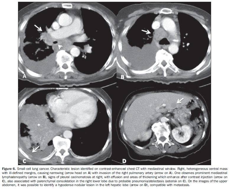
OBJECTIVE: To describe key imaging findings in a series of cases of primary neuroendocrine tumors of the lung (NTLs), with emphasis on computed tomography changes. MATERIALS AND METHODS: Imaging studies of 22 patients (12 men, mean age 60 years) with histopathologically confirmed diagnosis, evaluated in the authors's institution during the last five years were retrospectively reviewed by two radiologists, with findings being consensually described focusing on changes observed at computed tomography. RESULTS: The authors have described five typical carcinoids, three atypical carcinoids, three large-cell neuroendocrine carcinomas (LCNCs), and 11 small-cell lung cancers (SCLCs). Only one typical carcinoid presented the characteristic appearance of central endobronchial nodule with distal pulmonary atelectasis, while the others were pulmonary nodules or masses. The atypical carcinoids corresponded to peripheral heterogeneous masses. One out of the three LCNCs was a peripheral homogeneous mass, while the others were ill-defined and heterogeneous. The 11 SCLCs corresponded to central, infiltrating and heterogeneous masses with secondary pleuropulmonary changes. Calcifications were absent both in LGNCs and SCLCs. Metastases were found initially and also at follow-up of all the cases of LCNCs and SCLCs. CONCLUSION: Although some imaging features may be similar, radiologic findings considered together with clinical information may play a relevant role in the differentiation of histological types of NTLs.
Computed tomography; Lung neoplasms; Neuroendocrine tumors


