Abstract
Objective:
To describe and quantify the main changes seen on computed tomography of the chest in mildly symptomatic adult patients with sickle cell disease, as well as to evaluate the radiologist accuracy in determining the type of hemoglobinopathy.
Materials and Methods:
A prospective study involving 44 adult patients with sickle cell disease who underwent inspiration and expiration computed tomography of the chest. The frequency of tomography findings and the extent of involvement are reported. We also calculated radiologist accuracy in determining the type of hemoglobinopathy by analyzing the pulmonary alterations and morphology of the spleen.
Results:
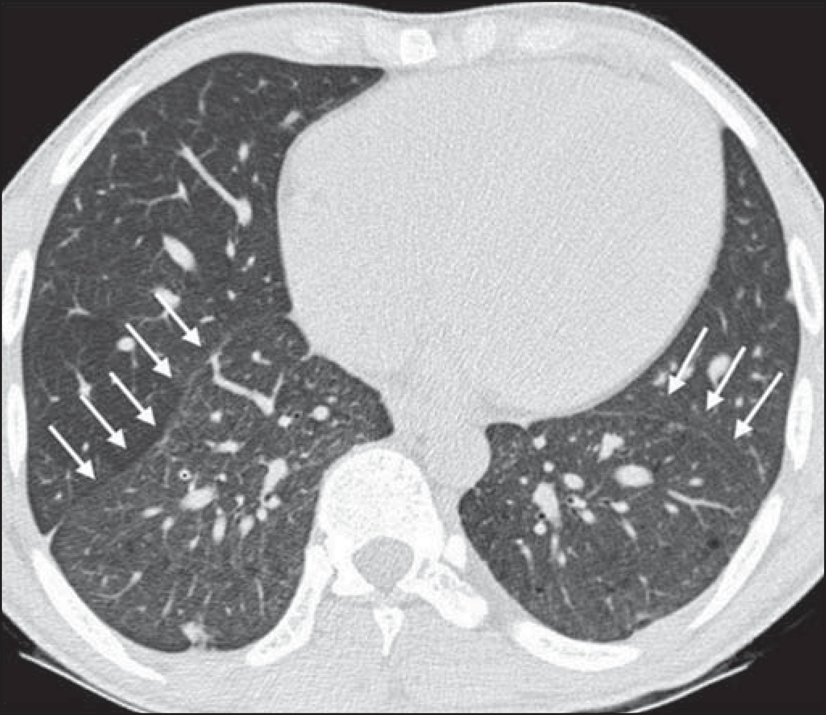
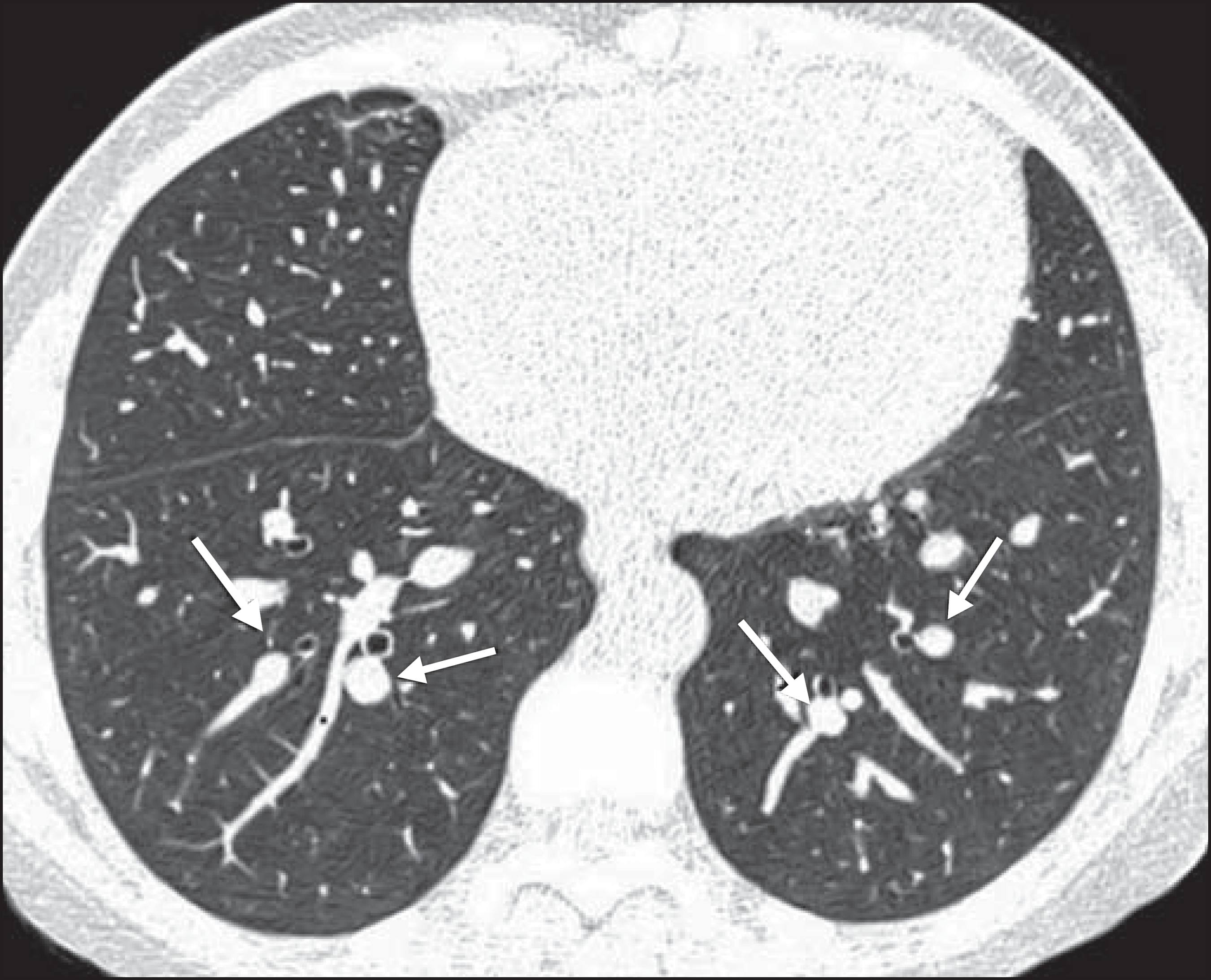
The changes found on computed tomography scans, in descending order of frequency, were as follows: fibrotic opacities (81.8%); mosaic attenuation (56.8%); architectural distortion (31.8%); cardiomegaly (25.0%); lobar volume reduction (18.2%); and increased caliber of peripheral pulmonary arteries (9.1%). For most of the findings, the involvement was considered mild, five or fewer lung segments being affected. The accuracy in determining the type of hemoglobinopathy (HbSS group versus not HbSS group) was 72.7%.
Conclusion:
In adult patients with sickle cell disease, the main tomography findings reflect fibrotic changes. In addition, computed tomography can be helpful in differentiating among hemoglobinopathies.
Keywords:
Anemia, sickle cell; Lung diseases/etiology; Tomography, X-ray computed