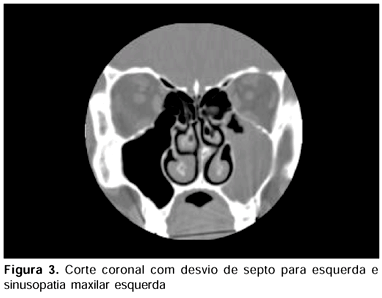
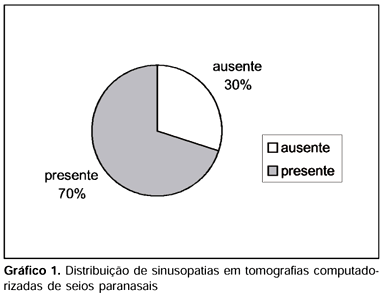
Introduction: Computed tomography has been increasingly used both to identify and to evaluate anatomy variations of nasal cavities that can lead to the development of sinusitis. Purpose: The purpose of the present study is to determine the incidence of mucosal abnormalities in paranasal sinuses found in CT scans of patients with symptons of sinusitis and analyze the correlation between sinusitis and presence of Haller's cell, concha bullosa and nasal septal deviation located in middle meatus. Study Design: Clinical retrospective. Material and Method: Paranasal sinus CT scans were obtained in 150 patients aged 13 years or more, from July 1999 to October 2001. The CT scans were performed in the Department of Radiology of Universidade Federal de São Paulo - Escola Paulista de Medicina. Patients with history of skull base or sinus surgery and tumor in these regions were excluded. Results: 70% of patients present mucosal abnormalities at least in one paranasal sinus. Maxillary sinusitis were observed in 52,7% of sinus, ethmoidal sinusitis in 28,0%, sphenoidal sinusitis in 13,0% and frontal sinusitis in 8,3%. Concha bullosa was observed in 33,3% of nasal cavities, nasal septal deviation (located in middle meatus) in 23,3% and Haller's cell in 9,3%. Conclusions: The most affected paranasal sinuses were: maxillary, ethmoid, sphenoid and frontal. Correlation between sinusitis and presence of Haller's cell, concha bullosa and nasal septal deviation (located in middle meatus) was not observed.
anatomy; nasal cavity; turbinates; paranasal sinuses; tomography


