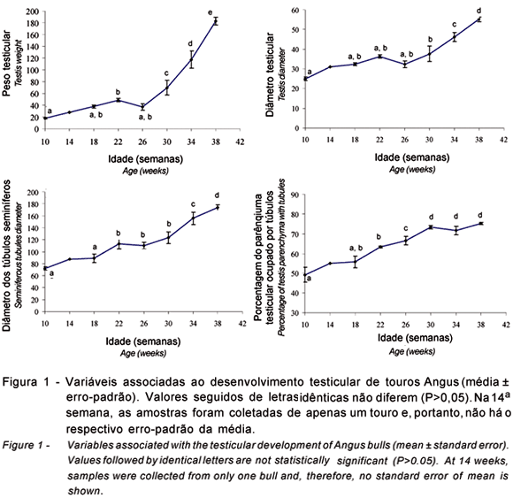
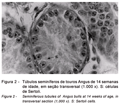
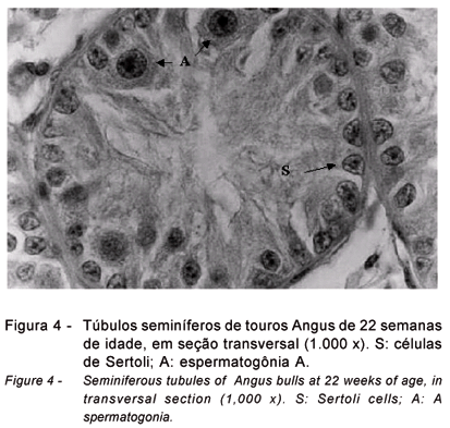
This study aimed to evaluate changes in hormone secretion and in seminiferous epithelium of Angus bulls between 10 and 38 weeks of age. Samples of testicular parenchyma and blood were collected from 25 animals castrated in 4 week intervals. Traits associated to testicular development and quantitative aspects of spermatogenesis and hormonal concentrations were transformed by logarithm before analyses of variance. Changes in testis and seminiferous tubule diameter and testis weight were more pronounced after 26 weeks of age. The percentage of testicular parenchyma occupied by seminiferous tubules increased from 49.3 to 75.2% from 10 to 38 weeks. Most tubules (>90%) had only Sertoli cells at 10 and 14 weeks, but the number of tubules with gonocytes and A spermatogonia increased at 18 (13.8±1.7%) and 22 weeks (19±1%). Tubules with B and intermediate spermatogonia became predominant at 26 weeks (24.5±8.2%) and those with spermatocytes as the most advanced germ cell type were more evident at 30 weeks (42.3±9.9%). Round spermatids were detected at 26 weeks and at 38 weeks of age, 62.3±1.5% of all tubules had either elongate or mature spermatids. Variations in testis growth (specially testis weight) after 26 weeks were coincident with the establishment of meiosis in the seminiferous tubules, morphological alterations in nucleus and nucleolus of the Sertoli cells (indicators of Sertoli cell differentiation), lower levels of androstenedione and significant increases in testosterone and estradiol 17beta. Associations between testis development and concentrations of FSH and LH were less evident.
estradiol; Sertoli; testis; testosterone


