INTRODUCTION:
Within the dental clinic blocking the inferior alveolar nerve is the most commonly used, however, several studies have shown higher rates of failure usually have reason to fail in observing the position of the shape and anatomy of the nerves. These failures resulting from anatomic variations of the inferior alveolar nerve have been studied in the literature from studies with the analysis of panoramic radiographs as proposed in this study.
OBJECTIVE:
The aim of this study was to investigate the occurrence and prevalence of anatomical variations as well as the correlated movements of the jaw with the canals side and sex that may occur in the mandibular canal in panoramic radiographs of patients of the Department of Dentistry, UFS.
MATERIAL AND METHOD:
Fifteen hundred panoramic radiographs of patients enrolled in the Department of Dentistry, Federal University of Sergipe (UFS) were analyzed radiographic images were observed on a light box using a black mask around the radiographs in an environment with proper lighting.
RESULT:
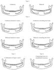
In this study 5.3% of bifurcations of the mandibular canal were observed, 47.5% of high canals, 16.8% of intermediate canals, 27.1% to 8.6% lower canals and canals with other variations.
CONCLUSION:
Based on the height of the mandibular canal was more prevalent among higher canals than other women, and there were no gender differences with respect to other types and affected sides. In the classification of bifid canals there was no statistically significant difference between men and women, the highest prevalence was for canals without bifurcation.
Mandibular nerve; oral surgery; panoramic radiography

 Thumbnail
