Abstract
The freshwater copepod Odontodiaptomus thomseni (Brehm, 1933) (Calanoida: Diaptomidae) is a rare species that has been reported only once - in its original description (BREHM 1933). The lack of subsequent records led to its inclusion in the Red List of threatened species (IUCN). Here we present a new record for O.thomseni. It was discovered in Salto Grande reservoir, which is located in the lower stretches of the Uruguay River, between Uruguay and Argentina, at the River Plate basin. In January 2010, three specimens (two males and one female) were found, and these were studied in detail using scanning electron microscopy (SEM). We only had material of Odontodiaptomus paulistanus (Wright, 1936) for comparison, but the position of the lateral spine in right P5 of the male, and the shape and size of lateral wings of the female are especially distinctive. Odontodiaptomus thomseni remains a rare species and we recommend keeping it on the IUCN Red List.
Copepod; rare; red list; taxonomy
TAXONOMY AND NOMENCLATURE
Rediscovered after 77 years: Odontodiaptomus thomseni a rare species of calanoid (Crustacea: Copepoda) from South America
Gilmar Perbiche-NevesI; Geoff A. BoxshallII; Carlos E. F. da RochaI; Marcos G. NogueiraIII
IDepartamento de Zoologia, Instituto de Biociências, Universidade de São Paulo. Rua do Matão, Travessa 14, 321, 05508-900 São Paulo, SP, Brazil. Email: gilmarpneves@yahoo.com.br
IIDepartment of Zoology, The Natural History Museum. Cromwell Road, London SW7 5BD, United Kingdom
IIIInstituto de Biociências, Universidade Estadual Paulista. Rubião Junior, 18618-000 Botucatu, SP, Brazil
ABSTRACT
The freshwater copepod Odontodiaptomus thomseni (Brehm, 1933) (Calanoida: Diaptomidae) is a rare species that has been reported only once in its original description (BREHM 1933). The lack of subsequent records led to its inclusion in the Red List of threatened species (IUCN). Here we present a new record for O.thomseni. It was discovered in Salto Grande reservoir, which is located in the lower stretches of the Uruguay River, between Uruguay and Argentina, at the River Plate basin. In January 2010, three specimens (two males and one female) were found, and these were studied in detail using scanning electron microscopy (SEM). We only had material of Odontodiaptomus paulistanus (Wright, 1936) for comparison, but the position of the lateral spine in right P5 of the male, and the shape and size of lateral wings of the female are especially distinctive. Odontodiaptomus thomseni remains a rare species and we recommend keeping it on the IUCN Red List.
KEY WORDS: Copepod; rare; red list; taxonomy.
The freshwater calanoid copepod fauna of the Neotropical region has many species. Currently 18 genera and 82 valid species in Diaptomidae are recorded from the Neotropics (BOXSHALL & HALSEY 2004), most of which are endemic to South America. The geographical distribution of those species is typically restricted by hydrographic basins and latitudinal ranges (BOXSHALL & DEFAYE 2008, SANTOS-SILVA 2008).
Odontodiaptomus was established by KIEFER (1936) and it currently consists of only three species: Odontodiaptomus thomseni (Brehm, 1933) (the type species), Odontodiaptomus michaelseni (Mrázek, 1901) and Odontodiaptomus paulistanus (Wright, 1936). Odontodiaptomus paulistanus has been recorded many times and appears to be widely distributed across the eastern region of the state of São Paulo State in Brazil, with its western limit in the floodplain of the Paraná River, state of Paraná (LANSAC-TÔHA et al. 2009), its southern limit at the Segredo Reservoir on the Iguaçu River (LOPES et al.1997), and its northern limit at the city of Juiz de Fora in the the state of Minas Gerais (WRIGHT 1936, 1937). Odontodiaptomus michaelseni has been recorded from across Uruguay and Argentina in areas that are close to the mouth of the Plate River, around the confluence of the Paraná and the Uruguay rivers. By contrast, the only valid record for O. thomseni is that of its original description by BREHM (1933), from Uruguay. REID (1996) included this species in the Red List of threatened species (IUCN Red List of Threatened Species, 2010) due to the lack of recent records and lack of data on it.
The rediscovery of the species after 77 years is documented here and a redescription, including a detailed taxonomic diagnosis and new images using scanning electron microscopy (SEM) are presented.
MATERIAL AND METHODS
Material was collected on 28 February, 2010, from two sites in the Salto Grande reservoir, located on the border with Uruguay (Salto Province) and Argentina (Entre Rios Province). The sampling sites (Fig. 1) are adjacent to the dam (31º15'31.41"S, 57º55'33.66"W) and upstream from the dam (30º46'27.52"S, 57º47'55.53"W).
Limnological variables were measured during sampling (Tab. I). Plankton samples were collected by vertical hauls using a conical plankton net of 68 µm mesh size, equipped with an antirefluxing device. The samples were preserved using Karnovski's solution (glutaraldehyde 2.5%, paraformaldehyde 20%).
The copepods were sorted and identified under a stereo-microscope and three individuals of Odontodiaptomus thomseni were found, two males and one female. One female and one male were used for SEM, the remaining male has been retained in the collection of the first author.
The preparation of specimens for SEM investigation followed the protocols of FELGENHAUER (1987) and HUYS & BOXSHALL (1991). The material was washed in 0.1M phosphate buffer solution at pH 7.3 (3 changes, five minutes each), then preserved in Osmium tetroxide (0.5% in water) for 20 minutes. The material was dehydrated through graded ethanol as follows: 7.5%, 15%, 30% and 50% (two changes in each concentration, five minutes); after 70% (three changes, 10 minutes); and to 90% and 100% (two changes in each concentration, five minutes). Then the material was stored in permeable plastic capsules and critical-point-dried in a BALZERD UNION CTD-020, using Carbon dioxide as the exchange medium). Finally the copepods were mounted on stubs. At this point some limbs were dissected off the female using fine insect pins to provide the best view of particular limbs; the male was kept intact. The material was sputter-coated with 15 nm of gold (BALZERS UNION MED-10) and analyzed using a Quanta 200 FEI scanning electronic microscope.
The taxonomic diagnosis presented here supplements and updates that of BREHM (1933), using SEM images as well as light microscope observations. Morphological terminology follows SANTOS-SILVA et al. (1999) and PAGGI (2006).
TAXONOMY
All observations were compared with a second species of the genus, O. paulistanus, using material collected by Carlos E.F. da Rocha in the Ecological Station of Boracéia, São Paulo State, Brazil.
Odontodiaptomus thomseni (Brehm, 1933) Figs 1-31
"Diaptomus" thomseni Brehm, 1933, p. 221, figs 1-4 ; Wright, 1937: 83, plate 1, fig. 8 , plate 2, fig. 1.
Diagnosis. Male: prosome with patches of setules on dorsal surface (Fig. 3 ); with sclerotized tooth-like process present on left caudal ramus, and other similar processes on surface of caudal ramus and urosome (Figs 4 and 5 ); two vestigial setae present on segment 2 of right geniculate antennule; well-developed modified setae present on segments 10, 11 and 13 of right antennule (Fig. 10 ); hyaline lamella on segment 20 of right antennule; endopod of right leg 5 well developed, 2-segmented (Figs 7 and 8 ); lateral spine of second exopod segment of right leg 5 positioned basally on segment and reaching beyond middle of segment (Fig. 7 ); terminal claw longer than segment (Fig. 7 ). Female: patches of setules present on dorsal surface of prosome; posterolateral wings of prosome strongly asymmetrical (Fig. 25); left lateral wing of genital double-somite with tapering process bearing spine at apex (Fig. 25); second urosomite with wing-like expansion on right side (Fig. 25); sclerotized tooth-like process on left side of third urosomite (Fig. 26); endopod of leg 5 well developed (Fig. 29).
Redescription. Male. Prosome (Fig. 2 ): Body length 989 µm, mean width 252 µm. Posterolateral corners of prosome not produced into wing-like expansions; suture between pedigerous somites 4 and 5 complete; groups of long, fine spinules present on dorsal and lateral surfaces of pedigerous somites 3 to 5 (Fig. 3 ). Urosome: 5-segmented; genital somite symmetrical. Caudal rami (Figs 2, 4 and 5 ): Semicircular sclerotized processes present on inner distal margin of left caudal ramus; also with some other sclerotized processes on dorsal, ventral and lateral surfaces of left caudal ramus (Figs 4 and 5 ) and also in different places. Inner margins of rami with setule rows. Right antennule (Figs 6 , 14 and 15 .): 22-segmented, geniculate, and swollen in mid section; numbers of setae (s), conical setae (cs), long setae (Ls), modified setae (ms), vestigial setae (vs), aesthetascs (ae) and spiny processes (p) on each segment as follows: (1) 1s + 1ae, (2) 2vs + 3s + 1ae, (3) 1vs +1Ls + 1ae, (4) 1s, (5) 1Ls + 1vs +1ae, (6) 1s, (7) 1Ls + 1ae, (8) 1s + 1cs, (9) 1s + 1Ls, (10) 1ms + 1s, (11) 1ms + 1s, (12) 2s + 1ae, (13) 1ms + 1s + 1ae, (14) 1s + 1Ls + 1ae, (15) 1ms + 1Ls + 1p + 1ae, (16) 1ms + 1Ls + 1p + 1ae, (17) 1s + 2ms, (18) 2ms + 1s, (19) 3ms + 1Ls + 1ae, (20) 2s + 2Ls + 1p, (21) 2Ls, (22) 4s + 1ae. Second segment with groove indicating compound origin. Spinous processes on segments 10 and 11 well developed, but smaller than that
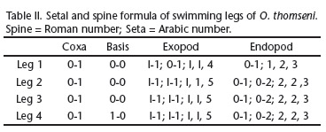
on segment 13. Segment 14 without spinous process; segments 15 and 16 each with medium-size spine. Modified setae on segments 15 and 16 rhomboidal. Segment 20 with well-developed denticulate hyaline membrane, denticles tiny proximally becoming more tooth-like distally. Left antennule: 25-segmented, number of setae (s), conical setae (cs), long setae (Ls), vestigial setae (vs) and aesthetascs (ae) on each segment as follows: (segment 1) 1s + 1ae, (2) 3s + 1ae +1vs, (3) 1Ls + 1ae + 1vs, (4) 1s, (5) 1Ls + 1ae +1vs, (6) 1s + 1vs, (7) 1Ls + 1ae, (8) 1s + 1cs, (9) 1s + 1Ls + 1ae, (10) -, (11) 1Ls, (12) 1s + 1cs + 1ae, (13) 1s, (14) 1Ls + 1ae, (15) 1Ls, (16) 1Ls + 1ae, (17) 1s, (18) 1Ls, (19) 1s + 1ae, (20) 1s, (21) 1Ls, (22) 1s + 1Ls, (23) 1Ls + 1ae, (24) 2Ls, (25) 1s + 3Ls + 1ae. Similar to female at Figs 20-22 and 27. Swimming legs (Figs 11 and 12): Exopods 3-segmented; endopods 3-segmented in legs 2-4 and 2-segmented in leg 1; Schmeil´s organ present on second endopod segment of leg 2; endopods of legs 1-4 with row of 4-8 spinules along distal margin; exopods of legs 1 to 4 with 3-5 denticles distally; row of setules present on basis of leg 1. Spine and seta formula of swimming legs as in Tab. II. Similar to female at Fig. 24. Right leg 5 (Figs 7-9 , 13 and 16 ): Coxa with small rounded process carrying apical spine located mid-distally on anterior surface (Fig. 16 ). Basis ca. 1.3 longer than wide, with rounded process on inner margin and seta on outer margin (Fig. 16 ). Endopod 2-segmented, length about 2/3 length of lateral spine on second exopod segment; ornamented with fringe of spinules around apex (Figs 7, 8 , 13 and 16 ). Exopod 3-segmented; with large circular projection larger than segment itself, located distally on mid-posterior surface of first segment (Fig. 16 ). Second segment of exopod ca. 2.2 longer than wide (Figs 7 and 16 ); lateral spine inserted near base of segment, lateral spine more than half as long as segment (Figs 7 , 13 and 16 ). Terminal claw incorporating third exopodal segment, 1.5 times longer than second segment, row of denticles ventrally, larger near base but decreasing in size distally (Figs 7, 9 and 16 ). Left leg 5 (Fig. 16 ): Coxa about as long as wide, with conspicuous sclerotized process on outer distal margin. Basis ca. 1.3 longer than wide with seta inserted midway along outer margin, seta about as long as width of segment. Exopod 2-segmented; first segment with curved outer margin, inner margin with rounded process ornamented with setules; second segment ending in digitiform process; inner margin curved; carrying stout spine. Endopod 1-segmented, about half as long as endopod of right leg; ending in row of spinules on distal margin.
Female. Prosome: Body length: 1,662 µm, maximum width: 283 µm. Slightly asymmetrical rostrum, with paired rostral filaments but right thickened in their median part (Fig. 17). Suture between fourth and fifth pedigerous somites incomplete; groups of long, fine spinules present on dorsal and lateral surfaces of pedigerous somites 3 to 5. Fifth pedigerous somite with well-developed posterolateral wings (Fig. 25); wings asymmetrical, right larger than left and terminating in sharp spinous process, with two chitinuous protuberances. Urosome: Genital double-somite asymmetrical, ca. 2 broader than longer; with well-developed spinous process on left side, at tip of expansion (Fig. 25); right side with large curved expansion on outer margin; genital area protruding; anterior part as a wide cuticular pad, with a crescentic margin; posterior pad with straight margin; gonoporal plates oval, symmetrical, showing the gonoporal slits. Part posterior to genital area showing some transverse cuticular folds (Fig. 23); second urosomite as wide as long (Fig. 25), with rounded chitinous process on margin of left side, plus other smaller processes on dorsal and ventral surfaces of segment (Fig. 26). Caudal rami: Symmetrical, 1.5 times longer than wide, ornamented with setules along inner and outer margins of both caudal rami; caudal setae thick. Antennula: Antennules reaching end of caudal rami. Setal formula as for left antennule of male (Figs 20-22 and 27). Antenna (Figs 18 and 19): Biramous, coxa with one seta, basis with two setae. Endopod 2-segmented: first segment with two setae in mid part; ornamented with row of spinules (between 4-5) distally and pore near row: compound distal segment with 15 setae, inner group of 8 setae plus distal group of 7 setae; row of 8-10 spinules present near distal margin. Exopod 7-segmented, setal formula: 1, 3, 1, 1, 1, 1, 4; segment 2 partially subdivided by suture on one side. Swimming legs (Fig. 24 5H): similar to male. Leg 5: Leg 5 symmetrical (Figs 28-31); coxa with process at outer distal corner bearing triangular sensilla at tip; basis with curved outer margin,and bearing long seta extending to tip of first exopod segment. Endopod 2- segmented (Fig. 29); first segment larger than second, almost 2.5 times longer than wide; second segment with two setae plus row of spinules at tip (Fig. 31). Exopod 3-segmented; first segment larger than second; second segment with lateral spine and with inner margin produced to form terminal claw ornamented with lateral rows of denticles along each side (Fig. 30); third segment small, offset and with two terminal setae, lateral smaller.
DISCUSSION
In his original work BREHM (1933) mentioned that he had found a "strange" copepod that was very different in morphology when compared with the known species. In fact, the species included in Odontodiaptomus share the possession of chitinous nodules on various parts of the body surface, including a toothlike process on the caudal rami and another on the urosome, from which the genus takes its name. Odontodiaptomus thomseni differs from the other two species included in Odontodiaptomus especially in the shape of the fifth leg of the male, which has the lateral spine on the second exopod segment inserted near the base of the segment, a well-developed endopod, and a rounded process on the first exopod segment. The females also differ markedly from the two other Odontodiaptomus species, particularly in the asymmetry of the genital receptacle. Consideration of these features makes it is easy to understand why BREHM (1933) regarded this species as "strange".
This is only the second confirmed record of O. thomseni. The discovery of only three individuals suggests the species israre and provides support for maintaining it on the Red List of threatened species. REID et al. (2002) pointed to possible factors that currently affect the Brazilian copepod fauna and impact species' abundance. These factors include the damming of rivers and disappearance of floodplain lakes; the introduction of alien species, and the increasing pollution of the rivers by domestic and industrial waste. Such factors may impact species abundances across the entire South America.
REID (1996) noted the existence of another record of O. thomseni from pools in Venezuela. We consider this record, so remote from the type locality (approximately 4,600 km), as not valid, without reexamination. BREHM (1933), WRIGHT (1938), MATSUMURA-TUNDISI (1986) and SANTOS-SILVA (2008) all emphasized that the three species of Odontodiaptomus are endemic to particular regions or latitudinal ranges, further casting doubt on the record from Venezuela. It is possible that the material reported from Venezuela represents a new species of the genus Odontodiaptomus. According to J.C. Paggi (pers. comm. 2011) there is another undescribed species of Odontodiaptomus inhabiting the Salto Grande reservoir (Uruguay/Argentina), although only one male individual has ever been found.
There are only three species of Odontodiaptomus and, as WRIGHT (1937) commented, O. thomseni is very different from O. paulistanus and O. michaelseni. These last two are very similar morphologically. In the absence of type material, is difficult to construct a dichotomous identification key due the paucity of knowledge of O. michaelseni. New material needs to be found near Buenos Aires.
Further study of this genus is necessary in order to fully understand its geographic distribution and taxonomic complexity. SUÁREZ-MORALES et al. (2005) point to the lack of information from some areas in the Neotropical region, and they show the distribution of Odontodiaptomus in South America as basically restricted to the La Plata River Basin.
More sampling should be concentrated in regions where O. thomseni has been found, especially in small water bodies, such pools and reservoirs. Although the nature of these small water bodies contrasts with the new locality reported here, this is the kind of environment sampled by BREHM (1933). Sampling such small habitats is important as PERBICHE-NEVES et al. (2011) demonstrated when they reported high abundance of Argyrodiaptomus bergi (Richard, 1898) in a pool at high altitude in the state of Santa Catarina, southern Brazil. Prior to this record, this copepod had not been found for 36 years.
ACKNOWLEDGEMENTS
The authors would like to thank FAPESP (process 2008/ 02015-7 and 2009/00014-6) for financial support to GPN; Danilo Naliato, José R. Debastiani Jr, and Silvia Casanova for help with fieldwork; and to the Electron Microscopy Centre (CME) of UNESP/Botucatu especially Tiago dos S. Tardivo for help with the acquisition of micrographic images.
LITERATURE CITED
Submitted: 14.XII.2011; Accepted: 27.III.2012.
Editorial responsibility: Marcos D.S. Tavares
- BOXSHALL, G.A. & S.H. HALSEY. 2004. An introduction to copepod diversity. London, The Ray Society, 966p.
- BOXSHALL, G.A. & D. DEFAYE. 2008. Global diversity of Copepods (Crustacea: Copepoda) in freshwater. Hydrobiologia 595: 195-207.
- BREHM, V. 1933. Diaptomus thomseni nov. spec., ein merkwürdiger neuer Diaptomus aus Uruguay. Zoologischer Anzeiger 104: 221-224.
- FELGENHAUER, B.E. 1987. Techniques for preparing crustaceans for scanning electron microscopy. Journal of Crustacean Biology 7: 71-76.
- HUYS, R. & G.A. BOXSHALL. 1991. Copepod evolution. London, The Ray Society, 468p.
- KIEFER, F. 1936. Über die Systematik der Südamerikanischen Diaptomiden (Crustacea Copepoda). Zoologischer Anzeiger 116: 194-200.
- LANSAC-TÔHA, F.A.; C.C. BONECKER; L.F.M. VELHO; N.R. SIMÕES; J.D. DIAS; G.M. ALVES & E.M. TAKAHASHI. 2009. Biodiversity of zooplankton communities in the Upper Paraná River floodplain: interannual variation from long-term studies. Brazilian Journal of Biology 69 (Suppl. 2): 539-549.
- LOPES, R.M.; F.A. LANSAC-TÔHA; R. VALE & M. SERAFIM-JÚNIOR. 1997. Comunidade zooplanctônica do Reservatório de Segredo, p. 39-60. In: A.A. AGOSTINHO & L.C. GOMES (Eds). Reservatório de Segredo: bases ecológicas para o manejo. Maringá, Eduem.
- MATSUMURA-TUNDISI, T. 1986. Latitudinal Distribution of Calanoida Copepods in Freshwater aquatic Systems of Brasil. Revista Brasileira de Biologia 46 (3): 527-553.
- PAGGI, J.C. 2006. Redescription and re-evaluation of the taxonomic status of the Neotropical copepod Diaptomus falcifer Daday, 1905 (Calanoida: Diaptomidae). Studies on Neotropical Fauna and Environment 41 (1): 67-78.
- PERBICHE-NEVES, G.; D. PREVIATTELLI & M.G. NOGUEIRA. 2011. Record of Argyrodiaptomus bergi (Crustacea: Copepoda: Calanoida) after 36 years and first record in Brazil. Zoologia 28 (5): 551-557.
- REID, J.W. 1996. Odontodiaptomus thomseni In: IUCN 2010. IUCN Red List of Threatened Species. Version 2010.4. Available online at: http://www.iucnredlist.org [Accessed: 15/XI/2010]
- REID, J.W.; I.E. BAYLY; G.L. PESCE; N.A. RAYNER; Y.R. REDDY; C.E.F. ROCHA; E. SUÁREZ-MORALES & H. UEDA. 2002. Conservation of continental copepod crustaceans, p. 253-261. In: E. ESCOBARBRIONES & F. ALVAREZ (Eds). Modern Approaches to study of Crustacea. Kluwer Academic, Plenum Publishers.
- SANTOS-SILVA, E.N. 2008. Calanoid of the families Diaptomidae, Pseudodiaptomidae, and Centropagidae from Brasil. Biologia Geral e Experimental 8 (1): 3-67.
- SANTOS-SILVA, E.N.; G.A. BOXSHALL & C.E.F. ROCHA. 1999. The neotropical genus Notodiaptomus Kiefer, 1936 (Calanoida: Diaptomidae): redescription of the type species Notodiaptomus deitersi (Poppe, 1891) and designation of a neotype. Studies on Neotropical Fauna and Environment 34: 114-128.
- SUÁREZ-MORALES, E.; J.W. REID & M. ELÍAS-GUTIÉRREZ. 2005. Diversity and distribuitional patterns of Neotropical Freshwater Copepods (Calanoida: Diaptomidae). International Review of Hydrobiology 90: 71-83.
- WRIGHT, S. 1936. Preliminary report on six new species of Diaptomus from Brasil. Anais da Academia Brasileira de Ciências 8: 79-85.
- WRIGHT, S. 1937. A review of some species of Diaptomus from São Paulo. Anais da Academia Brasileira de Ciências 9: 65-82.
- WRIGHT, S. 1938. A review of the Diaptomus bergi group, with descriptions of two new species. Transactions of the American Microscopical Society 57: 297-315.


Publication Dates
-
Publication in this collection
07 May 2012 -
Date of issue
Apr 2012
History
-
Received
14 Dec 2011 -
Accepted
27 Mar 2012