
Abstracts
The present research describes the histology of the midgut, gastric caeca, and pyloric valve of Tropidacris collaris (Stoll, 1813), (Orthopetera: Romaleidae). We used light microscopy, staining (Gomori's trichrome and periodic acid-Schiff (PAS)), and a routine histological analysis method (hematoxilin-eosin). The insects were obtained from, and also bred in, the Laboratory of Entomology, Department of Biology, of the Rural Federal University of Pernambuco (UFRPE). The collected material was fixed in alcoholic Boüin and embedded in paraplast. The results demonstrated that the midgut wall is composed of an inner epithelial layer and two outer layers of striate muscles: one internal (circular) and the other external (longitudinal), with connective tissue between the muscle fibers. The epithelium is single-layered, with two cell types: regenerative and elongated columnar. The gastric caeca presents muscle layers similar to those of the midgut. Simple columnar epithelium lines the gastric caeca, which presents villi and projects towards the lumen. The pyloric valve is of striate muscle tissue, covered by a single epithelial-cell layer.
histology; midgut; gastric caeca; pyloric valve; Tropidacris collaris
A presente pesquisa teve como objetivo descrever a histologia do mesêntero, cecos gástricos e válvula pilórica de Tropidacris collaris (Stoll, 1813) (Orthoptera: Romaleidae), por meio da microscopia de luz, utilizando-se colorações especiais (Tricrômico de Gomori e P.A.S. - Ácido periódico de Schiff) e de rotina (Hematoxilina-Eosina). Os insetos foram obtidos da criação existente no Laboratório de Entomologia, do Departamento de Biologia, da UFRPE. O material coletado foi fixado em Boüin alcoólico e processado para inclusão em "paralast". Os resultados mostraram que a parede do mesêntero é constituída por duas camadas de músculo estriado: uma interna (circular) e outra externa (longitudinal), com a presença de tecido conjuntivo entre as fibras musculares. O epitélio é do tipo simples, constituído por dois tipos de células: células regenerativas e células colunares altas. Os cecos gástricos também apresentam as camadas de músculo semelhantes às observadas no mesêntero. O tecido epitelial é do tipo simples colunar projetando-se para a luz do órgão, formando vilosidades. A válvula pilórica é constituída por tecido muscular estriado, recoberto por tecido epitelial.
histologia; mesêntero; cecos gástricos; válvula pilórica e Tropidacris collaris
BIOLOGY
Histological description of the midgut and the pyloric valve of Tropidacris collaris (Stoll, 1813) (Orthopetera: Romaleidae)
Descrição histológica do mesêntero e válvula pilórica de Tropidacris collaris (Stoll, 1813) (Orthoptera: Romaleidae)
Wanderley-Teixeira, V.I; Teixeira, A. A. C.I; Cunha, F. M.I; Costa, M. K. C. M.I; Veiga, A. F. S. L.II; Oliveira, J. V.III
IDepartamento de Morfologia e Fisiologia Animal, Universidade Federal Rural de Pernambuco, Rua Dom Manoel de Medeiros, s/n, CEP 52171-900, Recife, PE, Brazil
IIDepartamento de Biologia, Universidade Federal Rural de Pernambuco, Rua Dom Manoel de Medeiros, s/n, CEP 52171-900, Recife, PE, Brazil
IIIDepartamento de Agronomia, Universidade Federal Rural de Pernambuco, Rua Dom Manoel de Medeiros, s/n, CEP 52171-900, Recife, PE, Brazil
Correspondence to Correspondence to: Valéria Wanderley Teixeira Departamento de Morfologia e Fisiologia Animal Universidade Federal Rural de Pernambuco Rua Dom Manoel de Medeiros, s/n CEP 52171-900, Recife, PE, Brazil e-mail: vwt@bol.com.br
ABSTRACT
The present research describes the histology of the midgut, gastric caeca, and pyloric valve of Tropidacris collaris (Stoll, 1813), (Orthopetera: Romaleidae). We used light microscopy, staining (Gomori's trichrome and periodic acid-Schiff (PAS)), and a routine histological analysis method (hematoxilin-eosin). The insects were obtained from, and also bred in, the Laboratory of Entomology, Department of Biology, of the Rural Federal University of Pernambuco (UFRPE). The collected material was fixed in alcoholic Boüin and embedded in paraplast. The results demonstrated that the midgut wall is composed of an inner epithelial layer and two outer layers of striate muscles: one internal (circular) and the other external (longitudinal), with connective tissue between the muscle fibers. The epithelium is single-layered, with two cell types: regenerative and elongated columnar. The gastric caeca presents muscle layers similar to those of the midgut. Simple columnar epithelium lines the gastric caeca, which presents villi and projects towards the lumen. The pyloric valve is of striate muscle tissue, covered by a single epithelial-cell layer.
Keywords: histology, midgut, gastric caeca, pyloric valve, Tropidacris collaris.
RESUMO
A presente pesquisa teve como objetivo descrever a histologia do mesêntero, cecos gástricos e válvula pilórica de Tropidacris collaris (Stoll, 1813) (Orthoptera: Romaleidae), por meio da microscopia de luz, utilizando-se colorações especiais (Tricrômico de Gomori e P.A.S. Ácido periódico de Schiff) e de rotina (Hematoxilina-Eosina). Os insetos foram obtidos da criação existente no Laboratório de Entomologia, do Departamento de Biologia, da UFRPE. O material coletado foi fixado em Boüin alcoólico e processado para inclusão em "paralast". Os resultados mostraram que a parede do mesêntero é constituída por duas camadas de músculo estriado: uma interna (circular) e outra externa (longitudinal), com a presença de tecido conjuntivo entre as fibras musculares. O epitélio é do tipo simples, constituído por dois tipos de células: células regenerativas e células colunares altas. Os cecos gástricos também apresentam as camadas de músculo semelhantes às observadas no mesêntero. O tecido epitelial é do tipo simples colunar projetando-se para a luz do órgão, formando vilosidades. A válvula pilórica é constituída por tecido muscular estriado, recoberto por tecido epitelial.
Palavras-chave: histologia, mesêntero, cecos gástricos, válvula pilórica e Tropidacris collaris.
INTRODUCTION
The alimentary canal of insects is a tube, normally straight or coiled, which extends from the mouth to the anus, with three main sections having different embryonic origins: the foregut or stomodaeum; the midgut, made up of the ventriculus and gastric caeca; and the hindgut or proctodaeum (Uvarov, 1966; Borror & De Long, 1969; Belkin, 1976; Maranhão, 1976).
The midgut, which has different cell types, is the main organ of an insect's digestive tract, which also has an absorptive function (Cavalcante & Cruz-Landim, 1999). The gastric caeca contains bacteria and other digestive tube microorganisms that produce enzymes and vitamins. In addition, it is through this organ that water is absorbed and nutrients are digested (Gallo et al., 2002).
Since histological descriptions of insect digestive system are relatively few, markedly so in Brazil, the present research aimed to describe the histology of the mesenteron and the pyloric valve of Tropidacris collaris (Stoll, 1813) (Orthopetera: Romaleidae), a species that represents a serious economic threat due to the damage it wreaks in several species of wild palm trees and some useful plants, in order to elucidate the operation of its digestive system (Duraton et al., 1987).
MATERIAL AND METHODS
Adult insects of T. collaris were bred in and obtained from the Laboratory of Entomology, in the Department of Biology of the Rural Federal University of Pernambuco (UFRPE). The insects were anesthetized with ethylic ether and dissected under a model-1069 Quimis stereomicroscope. The collected material was immediately put in alcoholic Boüin and kept for two hours at room temperature. The material was then dissected, and returned to the fixing solution for four hours, following which it was placed in an 80% v/v solution of alcohol. The material was then dehydrated in two 15 min baths of absolute alcohol, and cleared in xylene for 15 min. After these procedures, the material was impregnated in Paraplast baths, diluted for 15 min at the following xylene percentages: 25%, 50%, 75%, and 100% and embedded after the last bath. Sections were then cut in Minot microtome (LEICA RM 2035) at 5 µm, and stained using a routine method (hematoxylin-eosin [HE]), and the special stains Gomori's trichrome, and periodic acid-Schiff (PAS). The morphological analysis of the material (photographed with an OLYMPUS BX-51 photomicroscope) was carried out using an OLYMPUS BX-49 light microscope.
RESULTS
The results showed that the mesenteron wall of T. collaris comprises two layers of striate muscles: internal (circular) with continuous fibers, and the external (longitudinal) separated by interstitial spaces (Fig. 1). These layers contained connective tissue between the muscle fibers (Fig. 2).
The single-layered columnar epithelium, which rests on a thin basal membrane, is constituted of two cell types: regenerative, with strongly acidic cytoplasm and a nucleus with dispersed chromatin, that are found in nests at the base of crypts formed by epithelium, and high columnar cells - the more common - found in the midgut, located close to the lumen, and the cells of which present microvilli. Connective tissue lies below the epithelium (Fig. 3). A peritrophic membrane is observed enveloping food particles (Fig. 4).
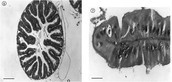
The histological section of the gastric caeca showed that its wall presents muscle layers similar to those of the alimentary canal. However, the epithelial tissue that rests on the basal membrane is columnar, projecting towards the lumen. This tissue forms villi, and its cells present microvilli (Figs. 5, 6, 7).
The constriction shown by a longitudinal section of the transition area between the mesenteron and the proctodaeum is the pyloric valve, made up of striate muscle tissue (sphincter) covered by simple epithelial tissue (Fig. 8).
DISCUSSION
The striated muscle tissue layers of the midgut are inverted with respect to those of the stomodaeum, which presents an external circular layer and an internal longitudinal one. This description is in agreement with that of several other authors (Borror & De Long, 1969; Duraton et al., 1987; Snodgrass, 1993; Gallo et al., 2002; Costa, 2003). The conjunctive tissue between muscle fibers and surrounding the muscles is connected to the peritoneal layer described by Buzzi & Miyazaki (1999) and Gallo et al. (2002).
According to Martoja & Ballan-Dufrançais (1984), Leite & Evangelista (2001), and Silva-Olivares et al., (2003), midgut epithelium is always single layered and composed basically of columnar, functional, and regenerative cells, thus confirming the results obtained in this research, which found that the epithelium of T. collaris is single-layered. However, those authors mention other types of cells, i.e., goblet, coprophilic, and those having an endocrine function. All of these may occur, in one or another species, or in one or another phase in the life cycle of any insect species.
Regenerative cells renew old cells or those functionally damaged. These cells may be scattered throughout the midgut, or maybe nested in crypts in the form of epithelial evaginations or papillae. The most common cell types in insect midguts is the columnar. Present in all insects, this cell has various names: principal, digestive, and enterocytic, and during its life cycle its functions are multiple: secretion of enzymes and glycoproteins, formation of the peritrophic membrane, nutrient absorption, and storage of organic and inorganic products. The luminal surface of these cells is generally covered by microvilli (Chapman, 1985; Borror et al., 1989; Cruz-Landim et al., 1996a, 1996b; Cavalcante & Cruz-Landim, 1999).
The epithelial disposition and presence of microvilli in the gastric caeca indicate an absorption area, which is in agreement with Gallo et al. (2002), who concluded that besides probably being involved in enteric bacteria storage and digestive enzyme secretion the gastric caeca also has a role in water and nutrient absorption.
The constriction observed in the pyloric valve, which regulates food passage to the proctodaeum, results from the projection of the muscular and epithelial layers towards the midgut lumen (Borror & De Long, 1969; Maranhão, 1976; Gallo et al., 1988; Snodgrass, 1993; Gallo et al., 2002).
In conclusion, the midgut wall is composed of an epithelial layer and striate muscles, with connective tissue between the muscle fibers. The simple epithelium contains both regenerative and columnar cells. The gastric caeca also present muscle layers similar to those of the midgut. Finally, the pyloric valve is composed of striate muscle tissue covered by a simple epithelial cell layer.
Received June 30, 2004
Accepted October 15, 2004
Distributed November 1, 2006
- BELKIN, J. N., 1976, Fundamentals of Entomology. Kinderhook: Ed. Biological Research Institute, pp. 24-25; 29-30.
- BORROR, D. J. & DE LONG, D. M., 1969, Introdução ao Estudo dos Insetos 1rst Ed. São Paulo: Edgard Blücher LTDA., pp. 19-22.
- BORROR, D. J., TRIPLEHORN, C. A. & JOHNSON, N. F. 1989, An Introduction to the Study of Insects. 6th Ed. Philadelphia: Saunders College Publishing, pp. 43-46.
- BUZZI, Z. J. & MIYAZAKI, R. D., 1999, Entomologia Didática 3rd Ed. Curitiba: Editora da UFPR, pp. 61-64.
- CAVALCANTE, V. M. & CRUZ-LANDIM, C., 1999, Types of cells present in the midgut of the insects: A Review. Naturalia, 24: 19-40.
- CHAPMAN, R. F., 1985, Coordination of digestion. In: Kerkut, G. A. & Gilbert, L. I. Comprehensive Insect Physiology, Biochemestry and Pharmacology. Oxford: Pergamon Press, 4: 213-240.
- COSTA, M. K. C. M. 2003, Descrição histológica dos principais constituintes do aparelho digestivo do gafanhoto Tropidacris Collaris (Stoll, 1813) (Orthoptera: Romaleidae) Monografia-Universidade Federal Rural de Pernambuco, 38p.
- CRUZ-LANDIM, C., SILVA-DE-MORAES, R. L. M., & SERRÃO, J. E. 1996a, Ultrastructural aspects of epithelial renewal in the midgut of adult worker bees (Hymenoptera: Apidae). J. Comp. Biol., 1:(1/2), 29-40.
- CRUZ-LANDIM, C., SERRÃO, J. E. & SILVA-DE-MORAES, R. L. M., 1996b, Cytoplasmic protrusion from digestive cells of bees. Cytobios, 88: 95-194.
- DURATON, J. F., LAUNOIS, M., LAUNOIS-LUONG, M. H. & LECOQ, M., 1987, Guia Prático de Luta Contra os Gafanhotos Devastadores no Brasil 1rst Ed. Roma: Fao-Cirad-Prifas, pp. 41-43.
- GALLO, D., NAKANO, O., NETO, S. S., CARVALHO, R. R. L., BATISTA, G. C., BERTI FILHO, E., PARRA, J. R. P., ZUCCHI, R. A., ALVES, S. B. & VENDRAMIM, I. O., 1988, Manual de Entomologia Agrícola 2nd Ed. São Paulo: Ceres, pp. 105-106.
- GALLO, D., MARCHINI, L. C., LOPES, J. R. S., & OMATO, C., 2002, Entomologia Agrícola 10th Ed. São Paulo: Ceres, pp. 133-138.
- LEITE, A. C. R. & EVANGELISTA, L. G., 2001, Ultrastructure of endocrine cells from the abdominal midgut epithelium of Lutzomyia longipalpis (Diptera: Psychodidae). Journal of Medical Entomology, 38(5): 749-752.
- MARANHÃO, Z. C. 1976, Entomologia Geral 3rd Ed. São Paulo: Nobel, pp. 145-154; 317-319.
- MARTOJA, R. & BALLAN-DUFRANÇAIS, C., 1984, The ultrastructure of digestive and excretory organs. In: King, R. C. & Akai, H. Insect Ultrastructure. 2º vol. 624p. New York: Plenum Press.
- SILVA-OLIVARES, A., DIAZ, E., SHIBAYAMA, M., TSUTSUMI, V., CISNEROS, R., & ZUIGA, G., 2003, Ultrastructural Study of the Midgut and Hindgut in Eight Species of the Genus Dendroctonus Erichson (Coleoptera: Scolytidae). Ann. Entomol. Soc. Am., 96(6): 883-900.
- SNODGRASS, R. E., 1993, Principles of Insect Morphology. 2nd Ed. London: Cornell University Press, pp. 359-374.
- UVAROV, S. B. 1966, Grasshoppers and Locusts. London: Cambridge University Press, pp. 70-89.
Correspondence to:
Publication Dates
-
Publication in this collection
13 Apr 2007 -
Date of issue
Nov 2006
History
-
Accepted
15 Oct 2004 -
Received
30 June 2004


