Abstract
Objectives:
Hemoglobin SC is the second most common variant of sickle-cell disease worldwide, after hemoglobin SS. The objectives of the study were to describe the clinical and laboratory characteristics of hemoglobin SC disease in children from a newborn screening program and treated at a blood center.
Methodology:
This study assessed a cohort of 461 infants born between 01/01/1999 and 12/31/2012 and followed-up until 12/31/2014. Clinical events were expressed as rates for 100 patient-years, with 95% confidence intervals. Kaplan-Meier survival curves were created.
Results:
The median age of patients was 9.2 years; 47.5% were female. Mean values of blood tests were: hemoglobin, 10.5 g/dL; reticulocytes, 3.4%; white blood cells, 11.24 × 10⁹/L; platelets, 337.1 × 10⁹/L; and fetal hemoglobin, 6.3%. Clinical events: acute splenic sequestration in 14.8%, blood transfusion 23.4%, overt stroke in 0.2%. The incidence of painful vaso-occlusive episodes was 51 (48.9-53.4) per 100 patient-years and that of infections, 62.2 episodes (59.8-64.8) per 100 patient-years. Transcranial Doppler ultrasonography (n = 71) was normal given the current reference values for SS patients. Hydroxyurea was given to ten children, all of whom improvement of painful crises. Retinopathy was observed in 20.3% of 59 children who underwent ophthalmoscopy. Avascular necrosis was detected in seven of 12 patients evaluated, predominantly in the left femur. Echocardiogram compatible with pulmonary hypertension was recorded in 4.6% of 130 children, with an estimated average systolic pulmonary artery pressure of 33.5 mmHg. The mortality rate from all causes was 4.3%.
Conclusions:
Clinical severity is variable in SC hemoglobinopathy. Several children have severe manifestations similar to those with SS disease.
KEYWORDS
Sickle-cell disease; Hemoglobinopathy SC; Children; Neonatal screening; Survival; Treatment
Resumo
Objetivos:
A hemoglobinopatia SC é a segunda variante mais comum da doença falciforme no mundo, após a hemoglobinopatia SS. Os objetivos do estudo foram descrever as características clínicas e laboratoriais da hemoglobinopatia SC em recém-nascidos diagnosticados por programa de triagem neonatal e encaminhados para acompanhamento em hemocentro.
Metodologia:
Coorte de 461 recém-nascidos SC nascidos entre 01/01/1999 e 31/12/2012 e seguidos até 31/12/2014. A incidência de eventos clínicos foi expressa por taxas relativas a 100 pacientes-ano, com limites de confiança a 95%. Curvas de sobrevida foram construídas segundo Kaplan-Meier.
Resultados:
Mediana de idade, 9,2 anos; 47,5%, feminino. Médias dos valores hematológicos: hemoglobina 10,5 g/dL; reticulócitos 3,4%; leucometria 11,24 x 10⁹/L; plaquetometria 337,1x10⁹/L; hemoglobina fetal 6,3%. Eventos clínicos: sequestro esplênico agudo em 14,8%, hemotransfusão 23,4%, AVC isquêmico 0,2%. A incidência de episódios vaso-oclusivos dolorosos foi de 51 (48,9-53,4) por 100 pacientes-ano; a de infecções, 62,2 episódios (59,8-64,8) por 100 pacientes-ano. Doppler transcraniano (n = 71) foi normal, se usados os valores de referência de crianças SS. Dez pacientes usaram hidroxiureia, todos com melhoria das crises dolorosas. Retinopatia foi observada em 20,3% das 59 crianças que fizeram fundoscopia. Necrose avascular foi detectada em 7 de 12 pacientes avaliados, com predomínio no fêmur esquerdo. Ecocardiograma compatível com hipertensão pulmonar foi registrado em 4,6% de 130 crianças, com média estimada de 33,5 mm Hg de pressão arterial pulmonar. A taxa de mortalidade por todas as causas foi de 4,3%.
Conclusões:
A hemoglobinopatia SC tem gravidade variável; várias crianças apresentam manifestações clínicas intensas, semelhantes às da hemoglobinopatia SS.
PALAVRAS-CHAVE
Doença falciforme; Hemoglobinopatia SC; Criança; Triagem neonatal; Sobrevida; Tratamento
Introduction
SC hemoglobinopathy is one of the most common variant of sickle-cell disease worldwide, second only to SS anemia. Hemoglobin C was first described in 1950 by Itano and Neel.11 Itano HA, Neel JV. A new inherited abnormality of human hemoglobin. Proc Natl Acad Sci USA. 1950;36:613-7. It is caused by a mutation in the sixth codon of the beta globin gene, which results in the substitution of glutamic acid by lysine (GAG > AAG).
The process that leads to clinical complications in individuals with SC hemoglobinopathy is not yet fully understood and appears to be related more to hyperviscosity and cell dehydration than to hemolysis-related vasculopathy, characteristic of SS disease.22 Nagel RL, Fabry ME, Steinberg MH. The paradox of hemoglobin SC disease. Blood Rev. 2003;17:167-78.
3 Lionnet F, Hammoudi N, Stojanovic KS, Avellino V, Grateau G, Girot R, et al. Hemoglobin sickle cell disease complications: a clinical study of 179 cases. Haematologica. 2012;97:1136-41.
4 Ballas SK, Lewis CN, Noone AM, Krasnow SH, Kamarulzaman E, Burka ER. Clinical, hematological, and biochemical features of Hb SC disease. Am J Hematol. 1982;13:37-51.-55 Pecker LH, Schaefer BA, Luchtman-Jones L. Knowledge insufficient: the management of haemoglobin SC disease. Br J Haematol. 2017;176:515-26.
The clinical events found in hemoglobin SS disease may also occur in patients with SC hemoglobinopathy.55 Pecker LH, Schaefer BA, Luchtman-Jones L. Knowledge insufficient: the management of haemoglobin SC disease. Br J Haematol. 2017;176:515-26.,66 Ayeroue J, Kafando E, Kam L, Gue E, Vertongen F, Ferster A, et al. Hemoglobin sickle cell disease: experience of the Yalgado Ouedraogo University Hospital of Ouagadougou, Burkina Faso. Arch Pediatr. 2009;16:316-21. However, the clinical manifestations appear to be less severe, except for proliferative retinopathy and necrosis of the femoral head.22 Nagel RL, Fabry ME, Steinberg MH. The paradox of hemoglobin SC disease. Blood Rev. 2003;17:167-78.,55 Pecker LH, Schaefer BA, Luchtman-Jones L. Knowledge insufficient: the management of haemoglobin SC disease. Br J Haematol. 2017;176:515-26.,66 Ayeroue J, Kafando E, Kam L, Gue E, Vertongen F, Ferster A, et al. Hemoglobin sickle cell disease: experience of the Yalgado Ouedraogo University Hospital of Ouagadougou, Burkina Faso. Arch Pediatr. 2009;16:316-21. Hydroxyurea seems to significantly reduce episodes of acute chest syndrome and painful vaso-occlusive crises (VOC) in children with SC hemoglobinopathy.22 Nagel RL, Fabry ME, Steinberg MH. The paradox of hemoglobin SC disease. Blood Rev. 2003;17:167-78.,55 Pecker LH, Schaefer BA, Luchtman-Jones L. Knowledge insufficient: the management of haemoglobin SC disease. Br J Haematol. 2017;176:515-26.,77 Yates AM, Dedeken L, Smeltzer MP, Lebensburger JD, Wang WC, Robitaille N. Hydroxyurea treatment of children with hemoglobin SC disease. Pediatr Blood Cancer. 2013;60:323-5.
8 Wang W, Brugnara C, Snyder C, Wynn L, Rogers Z, Kalinyak K, et al. The effects of hydroxycarbamide and magnesium on haemoglobin SC disease: results of the multi-centre CHAMPS trial. Br J Haematol. 2011;152:771-6.-99 Luchtman-Jones L, Pressel S, Hilliard L, Brown RC, Smith MG, Thompson AA, et al. Effects of hydroxyurea treatment for patients with hemoglobin SC disease. Am J Hematol. 2016;91:238-42.
The aim of this study was to describe the clinical and laboratory characteristics of hemoglobin SC disease in children from a neonatal screening program and followed at a blood center.
Methods
The descriptive study was based on a retrospective cohort, using the medical records from the blood center and the neonatal screening database. The population initially consisted of 569 children with FSC electrophoretic profile at birth, who were born between 01/01/1999 and 12/31/2012, screened by the Neonatal Screening Program of Minas Gerais (PTN-MG), and followed-up at the Hemocentro of Belo Horizonte (HBH) of Fundação Hemominas.
Twelve children were excluded from the analysis due to transfer to another blood center and 18 due to loss of clinical follow-up; 539 individuals were still eligible. Specific contact with the families for signing the free and informed consent form (FICF) was not possible in 78 cases.
The clinical and laboratory findings refer to the subtotal of 461 children included in the study (539 minus the 78 whose families did not sign the FICF). The evolution of these children was reviewed up to 12/31/2014, so that all the children were followed-up for a minimum of two years.
Clinical information, such as VOC, infections, acute splenic sequestration crisis (ASSC), priapism, ophthalmologic, orthopedic and cardiologic consultations, and acute cerebrovascular events were extracted from the medical records, as well as data on treatment (blood transfusions and hydroxyurea use).
A transcranial Doppler (TCD) was carried out in 71 children and interpreted by a single professional using Nicolet equipment (model EME TC 2000, Nicolet - Madison, WI, USA). A high risk of ischemic stroke was defined as time-averaged mean of the maximum velocity (TAMMX) > 200 cm/s in the internal carotid or middle cerebral arteries, according to the STOP1010 Adams RJ, McKie VC, Hsu L, Files B, Vichinsky E, Pegelow C, et al. Prevention of a first stroke by transfusions in children with sickle cell anemia and abnormal results on transcranial Doppler ultrasonography. N Engl J Med. 1998;339:5-11. protocol.
A complete blood count was performed using the Coulter T-890 equipment (Beckman Coulter, Inc., CA, USA). The reticulocytes were counted in smear stained with brilliant cresyl blue. All hematological values were transcribed from the medical records, in the absence of infectious processes or painful crises, and at least three months after the use of blood components. The arithmetic mean of hematological data was considered as the baseline value for each patient. The baseline fetal hemoglobin (FHb) was obtained through hemoglobin electrophoresis at the most advanced age within the follow-up period, provided that it was collected after two years of life.
In order not to overestimate mortality, the 78 children who did not have a signed FICF were included in the rate denominator, since they were alive according to the medical follow-up records.
The statistical analysis was performed using SPSS (IBM SPSS Statistics for Windows, Version 20.0. NY, USA). The results were expressed as mean and standard deviation (SD), or median and interquartile range, when the distribution of values was non-Gaussian. The incidence of ASSC, VOC, infections, and blood transfusions were expressed by rates relative to 100 patient-years, with 95% confidence intervals. The probability curves for the occurrence of the first episode of ASSC and death (Kaplan-Meier) were extracted from the "One minus Survival function" chart, which represents the function (1 - Survival function).
The study was approved by the Research Ethics Committee of the institution, Opinion 13327713.5.0000.5149. The patients and parents/guardians were asked to sign the FICF.
Results
Of the 461 children, 219 (47.5%) were girls. Age ranged from 1 to 16.7 years (median 9.2 years). At some point during the clinical course, 317 children (68.8%) had a palpable spleen below the left costal border, in the absence of an infectious process. The mean maximum values and the last spleen size value were 2.0 and 0.5 cm below the costal border, respectively. The mean ages when these values were measured were 5.8 years and 8.6 years, respectively.
At least one VOC was recorded in 344 patients (74.6%), and 21.9% of the cases were hospitalized. The time to the first VOC ranged from 2 months to 15 years (median 5.1 years). The incidence of VOC was 51 crises per 100 patient-years (95% CI: 48.9-53.4). Approximately 25% of the children did not present VOC during the study period.
The incidence of infectious episodes was 62.2 per 100 patient-years (95% CI: 59.8-64.8). Only 36 children (7.8%) did not have any infection recorded in the medical files. Upper airway infections were the most frequent (26.5%), followed by pneumonia/acute chest syndrome (17.2%), tonsillitis (14.6%), acute otitis media (8%), and fever of unknown origin (6.2%).
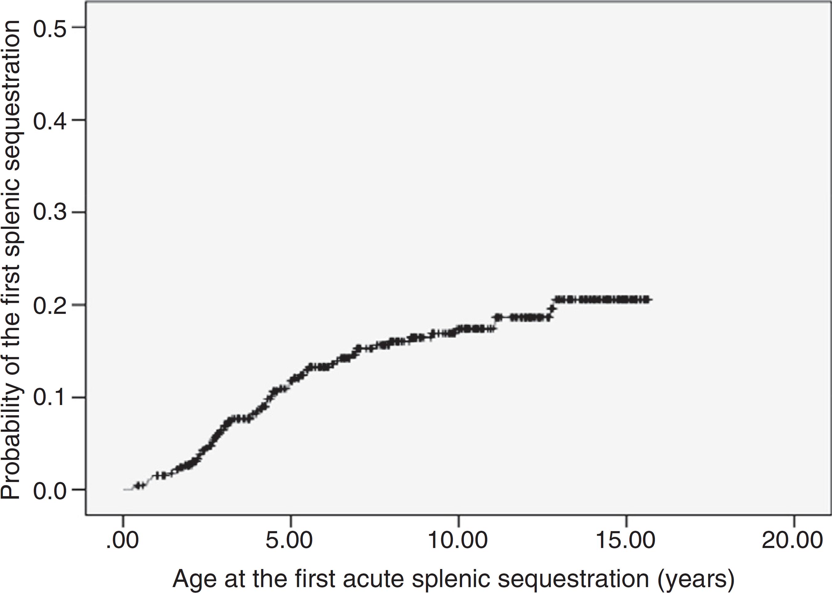
ASSC was recorded in 68 patients (14.8%), with recurrence rate of 25%. The incidence was of 1.9 first events per 100 patient-years (95% CI: 1.5-2.4). The age at the first event ranged from 3.5 months to 12.8 years (median 3.8 years). Most of the first episodes of ASSC (70%) occurred within the first five years of life. The probability curve for the occurrence of the first ASSC episode is shown in Fig. 1. Seven children underwent splenectomy, two after the first episode, three after the second, and one after the third. The age at the procedure ranged from 2.8 to 12.7 years (median 5.7 years).
Kaplan-Meier probability curve of the occurrence of the first episode of acute splenic sequestration. The estimated probability for the entire observation period was 20.6% (95% CI = 15.7-25.5%).
Four boys (0.9%) had priapism episodes. One patient had two events in less than 30 days, aged 7.9 years, while the other three had one event each, at 6.3, 7.3, and 9.7 years of age.
TCD was performed in 71 of the 461 children (15.4%), all with low risk of ischemic stroke (Table 1), using reference values for patients with SS hemoglobinopathy. One girl had ischemic stroke at age 11. Transthoracic and transesophageal echocardiographies were performed, which did not disclose intracardiac septal defects. The investigation of thrombophilia was negative. Cerebral magnetic resonance angiography evidenced bilateral vasculopathy, therefore being considered responsible for the cerebral ischemia. Sequencing of the HBB gene confirmed the S and C mutations and did not identify other mutations. The child had not undergone TCD before the stroke. At 15 years of age, TAMMX in the basilar artery was 113 cm/s, suggesting compensatory circulation secondary to stroke. Currently, the child has an adequate social behavior, but shows mild to moderate claudication in the lower left limb and moderate deficit in mathematical reasoning. She receives regular blood transfusions and psychopedagogical support.
Distribution of transcranial Doppler maximum mean velocities (cm/s) in 71 children with SC hemoglobinopathy.
A total of 232 children (50.3%) were referred to ophthalmologic assessment. Of this total, 59 (25.4%) underwent fundoscopy, and a finding compatible with retinopathy was verified in 20.3% of them. The median age at retinopathy diagnosis was 9.5 years. Regarding the orthopedic assessment, 12 children had a recorded medical consultation (2.6%), and avascular necrosis was observed in seven. Five showed a lesion in the left femur. One child had bilateral avascular necrosis in the shoulders and another had bilateral femoral lesions. As for the cardiologic assessment, 130 children (28.2%) underwent an echocardiogram during the follow-up period; 4.6% had findings suggestive of pulmonary hypertension, with an estimated mean of 33.5 mmHg PASP, which varied between 31 and 38 mmHg. No records of sickle cell nephropathy were retrieved from the files.
Regarding the clinical treatment, prescription of antimicrobial prophylaxis, folic acid supplementation, and immunization was recorded for all patients. It was not possible to adequately quantify treatment adherence, as there was no standardized recording of these data by the attending physicians.
Transfusion of packed red blood cells was necessary in 108 children (23.4%). As mentioned before, one patient received 63 transfusions as secondary ischemic stroke prevention. The other 107 patients received 185 transfusion procedures, with a mean incidence of 4.7 transfusions per 100 patient-years (95% CI: 4.0-5.4). They were caused by ASSC, preparation for surgical procedures, and clinical complications associated with infections. A total of 76.6% of the children did not receive transfusions; 15% received only one and 8.4%, two or more.
Ten (2.2%) children used hydroxyurea indicated due to repeated episodes of VOC in six cases. Avascular necrosis of the femoral head (one case) and ACS (three cases, alone or in combination with other events) comprised the other indications (Table 2). All had a significant reduction in the number of VOC. The child with bone necrosis showed marked gait improvement and had significant pain reduction.
Clinical and laboratory data of ten children with SC hemoglobinopathy who used hydroxyurea (HU).
The mean hemoglobin was 10.5 g/dL and the mean reticulocyte count was 3.4%. The distribution of hematological exams is shown in a summarized form in Table 3.
Hematologic exams in children with SC hemoglobinopathy treated in a Brazilian state Blood Center.
Among the 539 children initially enrolled for the study, there were 23 deaths (4.3%). In six cases it was not possible to determine the cause of death. Ten deaths were related to infectious complications, with six episodes of pneumonia/ACS and four episodes of sepsis. Of the seven remaining deaths, three occurred after ASSC. Acute aplastic crisis, gastroenteritis/dehydration, traumatic brain injury, and hemorrhagic shock were responsible for one death each. The age at death ranged from 3 months to 9.7 years, with a median of 3.2 years; 65.2% died younger than 5 years of age. The estimated probability of death until the age of 10 years was 5.4% (95% CI: 3.2-7.6%).
Discussion
The present study included 461 children with SC hemoglobinopathy. The severity of the clinical manifestations in SC disease varied from child to child. As shown by the maximum incidence rates of the different clinical events, several children had high values, suggesting a severity of the clinical course similar to that observed in SS children.
Approximately 75% of the patients had at least one VOC, a result similar to the 78% found by Tuttle and Koch,1111 Tuttle AH, Koch B. Clinical and hematological manifestations of hemoglobin CS disease in children. J Pediatr. 1960;56:331-42. but superior to that reported by Powars et al.,1212 Powars D, Chan LS, Schroeder WA. The variable expression of sickle cell disease is genetically determined. Semin Hematol. 1990;27:360-76. with 60% of cases. The incidence of VOC for the entire population in the present study was 51 crises per 100 patient-years. The American cohort had a mean incidence of 30 VOC per 100 patient-year.1313 Powars DR, Hiti A, Ramicone E, Johnson C, Chan L. Outcome in hemoglobin SC disease: a four-decade observational study of clinical, hematologic, and genetic factors. Am J Hematol. 2002;70:206-15. The same group observed that the time until the first report of pain in childhood was influenced by the disease genotype, occurring significantly earlier in SS disease (median of 13.9 months) than in SC disease (median of 43.6 months).1414 Dampier C, Ely B, Brodecki D, Coleman C, Aertker L, Sendecki JA, et al. Pain characteristics and age-related pain trajectories in infants and young children with sickle cell disease. Pediatr Blood Cancer. 2014;61:291-6. In the present study, the median was 61 months. Painful crises in children with SC hemoglobinopathy occur with approximately half the frequency of what is observed in sickle-cell anemia.22 Nagel RL, Fabry ME, Steinberg MH. The paradox of hemoglobin SC disease. Blood Rev. 2003;17:167-78.,55 Pecker LH, Schaefer BA, Luchtman-Jones L. Knowledge insufficient: the management of haemoglobin SC disease. Br J Haematol. 2017;176:515-26.,1212 Powars D, Chan LS, Schroeder WA. The variable expression of sickle cell disease is genetically determined. Semin Hematol. 1990;27:360-76.,1313 Powars DR, Hiti A, Ramicone E, Johnson C, Chan L. Outcome in hemoglobin SC disease: a four-decade observational study of clinical, hematologic, and genetic factors. Am J Hematol. 2002;70:206-15. However, it should be emphasized that even though the incidence is lower, it is relevant and should be considered by healthcare teams in emergency units, in order to avoid under-treatment and dismissal of pain complaints in patients with SC hemoglobinopathy.
Regarding the evaluation of target organs, a finding compatible with retinopathy was observed in 20.3% children submitted to fundoscopy. Most studies on retinopathy have been performed in adult patients, with a mean prevalence of approximately 33%.22 Nagel RL, Fabry ME, Steinberg MH. The paradox of hemoglobin SC disease. Blood Rev. 2003;17:167-78.,33 Lionnet F, Hammoudi N, Stojanovic KS, Avellino V, Grateau G, Girot R, et al. Hemoglobin sickle cell disease complications: a clinical study of 179 cases. Haematologica. 2012;97:1136-41.,1515 Elagouz M, Jyothi S, Gupta B, Sivaprasad S. Sickle cell disease and the eye: old and new concepts. Surv Ophthalmol. 2010;55:359-77. Among the studies that included children, the result was similar to that found by two North American groups, with 23% and 16.4%, respectively.1313 Powars DR, Hiti A, Ramicone E, Johnson C, Chan L. Outcome in hemoglobin SC disease: a four-decade observational study of clinical, hematologic, and genetic factors. Am J Hematol. 2002;70:206-15.,1616 Rosenberg JB, Hutcheson KA. Pediatric sickle cell retinopathy: correlation with clinical factors. J Aapos. 2011;15:49-53. Regarding the cardiological evaluation, 4.6% of the children who underwent echocardiography presented echocardiographic values that were suggestive of pulmonary hypertension, a result similar to that found in an English study,1717 Chaudry RA, Cikes M, Karu T, Hutchinson C, Ball S, Sutherland G, et al. Paediatric sickle cell disease: pulmonary hypertension but normal vascular resistance. Arch Dis Child. 2011;96:131-6. with 6%. However, what is noteworthy in the present study was the small number of children submitted to fundoscopy (25%) and echocardiogram (28.2%), since patients should be referred for annual ophthalmologic and cardiologic evaluations after the age of 5 years. It is possible that the actual incidence of sickle cell retinopathy and echocardiographic alterations suggestive of pulmonary hypertension are underestimated in the assessed population. This demonstrates the need to establish a permanent network of experienced interconsultants in proliferative retinopathy and pediatric cardiology for adequate early detection and treatment.
Regarding the orthopedic evaluation, a description of avascular necrosis was found in 58.3% of the assessed patients. In the literature, the prevalence varies between 8.8 and 15%,33 Lionnet F, Hammoudi N, Stojanovic KS, Avellino V, Grateau G, Girot R, et al. Hemoglobin sickle cell disease complications: a clinical study of 179 cases. Haematologica. 2012;97:1136-41.,1212 Powars D, Chan LS, Schroeder WA. The variable expression of sickle cell disease is genetically determined. Semin Hematol. 1990;27:360-76.,1313 Powars DR, Hiti A, Ramicone E, Johnson C, Chan L. Outcome in hemoglobin SC disease: a four-decade observational study of clinical, hematologic, and genetic factors. Am J Hematol. 2002;70:206-15.,1818 Yawn BP, Buchanan GR, Afenyi-Annan AN, Ballas SK, Hassell KL, James AH, et al. Management of sickle cell disease: summary of the 2014 evidence-based report by expert panel members. JAMA. 2014;312:1033-48. but the small number of cases in the present study hinders the establishment of a solid conclusion. Four boys (0.9%) presented priapism episodes during the follow-up period. Published data on priapism usually result from the joint analysis of the SS, SC, and S-beta thalassemia genotypes, with an approximate prevalence of 35% in adults.1919 Olujohungbe AB, Adeyoju A, Yardumian A, Akinyanju O, Morris J, Westerdale N, et al. A prospective diary study of stuttering priapism in adolescents and young men with sickle cell anemia: report of an international randomized control trial - the priapism in sickle cell study. J Androl. 2011;32:375-82. Likewise, the small number of cases and the retrospective design of the study limit the data analysis.
Sixty-eight children (14.8%) had ASSC. There are few studies on ASSC involving children with SC hemoglobinopathy; the prevalence is estimated to be between 5 and 12%.44 Ballas SK, Lewis CN, Noone AM, Krasnow SH, Kamarulzaman E, Burka ER. Clinical, hematological, and biochemical features of Hb SC disease. Am J Hematol. 1982;13:37-51.,2020 Serjeant GR, Ashcroft MT, Serjeant BE. The clinical features of haemoglobin SC disease in Jamaica. Br J Haematol. 1973;24:491-501.,2121 Aquino VM, Norvell JM, Buchanan GR. Acute splenic complications in children with sickle cell-hemoglobin C disease. J Pediatr. 1997;130:961-5. Most of the ASSC early episodes in the present study (70%) occurred within the first five years of life. A study performed by the same authors on 255 SS/β0 thalassemia children treated at the Belo Horizonte Blood Center showed that, in addition to the incidence of ASSC being approximately five times higher in children with SS/Sβ0 thalassemia (10.2 first episodes per 100 patient-years), 75% of them presented the first event before the age of 2 years.2222 Rezende PV, Viana MB, Murao M, Chaves AC, Ribeiro AC. Acute splenic sequestration in a cohort of children with sickle cell anemia. J Pediatr (Rio J). 2009;85:163-9. There was a convergence in the indication of splenectomy after two or more ASSC episodes in both studies. Most authors, however, recommend performing the surgical procedure after the first or, at most, after the second episode of ASSC.1818 Yawn BP, Buchanan GR, Afenyi-Annan AN, Ballas SK, Hassell KL, James AH, et al. Management of sickle cell disease: summary of the 2014 evidence-based report by expert panel members. JAMA. 2014;312:1033-48.,2121 Aquino VM, Norvell JM, Buchanan GR. Acute splenic complications in children with sickle cell-hemoglobin C disease. J Pediatr. 1997;130:961-5. The authors believe that, considering the presented data, the education of caregivers of children with SC disease in relation to spleen palpation is, therefore, as important as in children with SS disease.
Regarding baseline hematological laboratory tests (Table 3), the mean hemoglobin levels, white blood cells, reticulocyte counts and platelet counts were 10.5 g/dL, 11,236/mm3, 3.4% and 337.098/mm3, respectively. Anemia in SC hemoglobinopathy is generally milder than in SS patients, with mean hemoglobin values ranging from 10.5-11.5 g/dL.33 Lionnet F, Hammoudi N, Stojanovic KS, Avellino V, Grateau G, Girot R, et al. Hemoglobin sickle cell disease complications: a clinical study of 179 cases. Haematologica. 2012;97:1136-41.,2323 Dalibalta S, Ellory JC, Browning JA, Wilkins RJ, Rees DC, Gibson JS. Novel permeability characteristics of red blood cells from sickle cell patients heterozygous for HbS and HbC (HbSC genotype). Blood Cells Mol Dis. 2010;45:46-52. As previously described, reticulocytes are slightly increased, and platelet and leukocyte counts are within the normal range.22 Nagel RL, Fabry ME, Steinberg MH. The paradox of hemoglobin SC disease. Blood Rev. 2003;17:167-78.,55 Pecker LH, Schaefer BA, Luchtman-Jones L. Knowledge insufficient: the management of haemoglobin SC disease. Br J Haematol. 2017;176:515-26.
All children who underwent TCD showed a low risk of ischemic stroke (Table 1). It is known that the incidence of stroke is approximately four times lower than that in patients with SS hemoglobinopathy, with rates ranging between 0.8 and 3%.1212 Powars D, Chan LS, Schroeder WA. The variable expression of sickle cell disease is genetically determined. Semin Hematol. 1990;27:360-76.,2424 Ohene-Frempong K, Weiner SJ, Sleeper LA, Miller ST, Embury S, Moohr JW, et al. Cerebrovascular accidents in sickle cell disease: rates and risk factors. Blood. 1998;91:288-94. There are currently insufficient studies and data regarding the TAMMX reference values for patients with SC hemoglobinopathy, and the classification of patients with SS hemoglobinopathy is used in these cases.1818 Yawn BP, Buchanan GR, Afenyi-Annan AN, Ballas SK, Hassell KL, James AH, et al. Management of sickle cell disease: summary of the 2014 evidence-based report by expert panel members. JAMA. 2014;312:1033-48.,2424 Ohene-Frempong K, Weiner SJ, Sleeper LA, Miller ST, Embury S, Moohr JW, et al. Cerebrovascular accidents in sickle cell disease: rates and risk factors. Blood. 1998;91:288-94.,2525 Adams R, McKie V, Nichols F, Carl E, Zhang DL, McKie K, et al. The use of transcranial ultrasonography to predict stroke in sickle cell disease. N Engl J Med. 1992;326:605-10. Vieira et al.2626 Vieira C, de Oliveira CN, de Figueiredo LA, Santiago RP, Adanho CS, Santana SS, et al. Transcranial Doppler in hemoglobin SC disease. Pediatr Blood Cancer. 2017;64, http://dx.doi.org/10.1002/pbc.26342. Epub 2016 Dec 13.
http://dx.doi.org/10.1002/pbc.26342...
reported that 0.7% of exams presented a high risk of stroke in 558 children with SC hemoglobinopathy. These authors suggest that lower reference values should be used in children with SC to be characterized as at high risk for ischemic stroke, but this requires further studies.
One child in the present study had an ischemic stroke episode. Deane et al. described the case of a patient with SC hemoglobinopathy who had an ischemic stroke at the age of 5 years, considered secondary to the underlying disease after ruling out other possibilities; the TAMMX was 146 cm/s in the right middle cerebral artery and 126 cm/s in the left middle cerebral artery.2727 Deane C, Goss D, O'Driscoll S, Melllor S, Pohl K, Dick MC, et al. Transcranial Doppler scanning and the assessment of stroke risk in children with HbSC disease. Arch Dis Child. 2007;93:138-41. A case of stroke in a 7-year-old child was also described by Powars et al.2828 Powars D, Wilson B, Imbus C, Pegelow C, Allen J. The natural history of stroke in sickle cell disease. Am J Med. 1978;65:461-71. in 1978 and another in a 6-year-old Nigerian child by Lagunju and Brown2929 Lagunju IA, Brown BJ. Adverse neurological outcomes in Nigerian children with sickle cell disease. Int J Hematol. 2012;96:710-8. in 2012. Three pediatric cases were reported by Ohene-Frempong in a North-American collaboration study.2424 Ohene-Frempong K, Weiner SJ, Sleeper LA, Miller ST, Embury S, Moohr JW, et al. Cerebrovascular accidents in sickle cell disease: rates and risk factors. Blood. 1998;91:288-94. As previously mentioned, there are no current data on TAMMX values to perform stratify ischemic stroke risk in patients with SC hemoglobinopathy. Therefore, the TAMMX results of the present study (Table 1) contribute to fill some gaps in the understanding of cerebral vasculopathy of SC children.
The use of hydroxyurea caused significant symptom reduction, similar to what has been previously described.77 Yates AM, Dedeken L, Smeltzer MP, Lebensburger JD, Wang WC, Robitaille N. Hydroxyurea treatment of children with hemoglobin SC disease. Pediatr Blood Cancer. 2013;60:323-5.
8 Wang W, Brugnara C, Snyder C, Wynn L, Rogers Z, Kalinyak K, et al. The effects of hydroxycarbamide and magnesium on haemoglobin SC disease: results of the multi-centre CHAMPS trial. Br J Haematol. 2011;152:771-6.-99 Luchtman-Jones L, Pressel S, Hilliard L, Brown RC, Smith MG, Thompson AA, et al. Effects of hydroxyurea treatment for patients with hemoglobin SC disease. Am J Hematol. 2016;91:238-42. It was observed that no patient received the maximum dose of hydroxyurea (35 mg/kg/day) due to the supervenience of hematological toxicity. Luchtman-Jones et al. reported 133 children with SC hemoglobinopathy treated with hydroxyurea at 18 referral centers in the United States. Their study showed a 38% reduction in painful events after 12 months of drug use.99 Luchtman-Jones L, Pressel S, Hilliard L, Brown RC, Smith MG, Thompson AA, et al. Effects of hydroxyurea treatment for patients with hemoglobin SC disease. Am J Hematol. 2016;91:238-42.
The estimated probability of death at 10 years, from all causes, was 5.4%, well below the 13.7% rate recorded for children with SS/Sβ0 thalassemia and similar to the 4.4% recorded in a study that included the entire state of Minas Gerais, Brazil.3030 Sabarense AP, Lima GO, Silva LM, Viana MB. Survival of children with sickle cell disease in the comprehensive newborn screening programme in Minas Gerais, Brazil. Paediatr Int Child Health. 2015;35:329-32. The United States collaboration group reported a 9% mortality rate in a cohort of 284 patients.1313 Powars DR, Hiti A, Ramicone E, Johnson C, Chan L. Outcome in hemoglobin SC disease: a four-decade observational study of clinical, hematologic, and genetic factors. Am J Hematol. 2002;70:206-15. It is observed that most of the children (65.2%) died when aged younger than 5 years. This finding can be explained by the higher incidence, in the described age group, of severe acute events and high potential for lethality, such as infections and ASSC, the most frequent causes of death in the present study. A significant occurrence of deaths secondary to indeterminate causes (26%) was also observed, which corroborates the difficulties of health teams in identifying possible clinical complications secondary to sickle cell disease.
The main limitations of the present study include the retrospective analysis and the small amount of data on specialized consultations of the patients included in the study, which restricts the conclusions about the incidence of some clinical complications, such as proliferative retinopathy and necrosis of the head of long bones. The evaluation of the hydroxyurea effects also has sampling limitations. The main reason for the sample loss of 19% (108/569) in the clinical follow-up refers to the difficulty in collecting the signed FICF, which accounted for 72% of the losses. The analysis of epidemiological data such as gender, age, and number of visits recorded in the Blood Center charts does not indicate that these children would have a clinical course different from that of the 461 children included in the study, but this possibility cannot be ruled out. Although only children from the state of Minas Gerais were included in this study, it is possible that the results shown herein may be extrapolated to other states, since the C allele has a unicentric origin,22 Nagel RL, Fabry ME, Steinberg MH. The paradox of hemoglobin SC disease. Blood Rev. 2003;17:167-78. although other genetic and environmental factors may have a different effect on children from other states and countries.
In conclusion, the analysis of the results adds relevant information about the profile of the clinical and laboratory behavior of SC hemoglobin disease in Brazil, aiming at preventive and curative health actions. Acute complications and target-organ lesions occur at varying degrees and several children show intense clinical manifestations, similar to those observed in SS hemoglobinopathy.
-
☆
Please cite this article as: Rezende PV, Santos MV, Campos GF, Vieira LL, Souza MB, Belisário AR, et al. Clinical and hematological profile in a newborn cohort with hemoglobin SC. J Pediatr (Rio J). 2018;94:666-72.
-
FundingFundação Hemominas, Newborn Screening Program (Nupad-UFMG), Fundação de Amparo à Pesquisa de Minas Gerais (FAPEMIG), and Conselho Nacional de Desenvolvimento Científico e Tecnológico (CNPq).
Acknowledgements
To CNPq, Fapemig, and Nupad financial support to the project. To our colleagues from Fundação Hemominas for the logistical support. To the children and their families for the consent to perform the research and publication of the results.
References
-
1Itano HA, Neel JV. A new inherited abnormality of human hemoglobin. Proc Natl Acad Sci USA. 1950;36:613-7.
-
2Nagel RL, Fabry ME, Steinberg MH. The paradox of hemoglobin SC disease. Blood Rev. 2003;17:167-78.
-
3Lionnet F, Hammoudi N, Stojanovic KS, Avellino V, Grateau G, Girot R, et al. Hemoglobin sickle cell disease complications: a clinical study of 179 cases. Haematologica. 2012;97:1136-41.
-
4Ballas SK, Lewis CN, Noone AM, Krasnow SH, Kamarulzaman E, Burka ER. Clinical, hematological, and biochemical features of Hb SC disease. Am J Hematol. 1982;13:37-51.
-
5Pecker LH, Schaefer BA, Luchtman-Jones L. Knowledge insufficient: the management of haemoglobin SC disease. Br J Haematol. 2017;176:515-26.
-
6Ayeroue J, Kafando E, Kam L, Gue E, Vertongen F, Ferster A, et al. Hemoglobin sickle cell disease: experience of the Yalgado Ouedraogo University Hospital of Ouagadougou, Burkina Faso. Arch Pediatr. 2009;16:316-21.
-
7Yates AM, Dedeken L, Smeltzer MP, Lebensburger JD, Wang WC, Robitaille N. Hydroxyurea treatment of children with hemoglobin SC disease. Pediatr Blood Cancer. 2013;60:323-5.
-
8Wang W, Brugnara C, Snyder C, Wynn L, Rogers Z, Kalinyak K, et al. The effects of hydroxycarbamide and magnesium on haemoglobin SC disease: results of the multi-centre CHAMPS trial. Br J Haematol. 2011;152:771-6.
-
9Luchtman-Jones L, Pressel S, Hilliard L, Brown RC, Smith MG, Thompson AA, et al. Effects of hydroxyurea treatment for patients with hemoglobin SC disease. Am J Hematol. 2016;91:238-42.
-
10Adams RJ, McKie VC, Hsu L, Files B, Vichinsky E, Pegelow C, et al. Prevention of a first stroke by transfusions in children with sickle cell anemia and abnormal results on transcranial Doppler ultrasonography. N Engl J Med. 1998;339:5-11.
-
11Tuttle AH, Koch B. Clinical and hematological manifestations of hemoglobin CS disease in children. J Pediatr. 1960;56:331-42.
-
12Powars D, Chan LS, Schroeder WA. The variable expression of sickle cell disease is genetically determined. Semin Hematol. 1990;27:360-76.
-
13Powars DR, Hiti A, Ramicone E, Johnson C, Chan L. Outcome in hemoglobin SC disease: a four-decade observational study of clinical, hematologic, and genetic factors. Am J Hematol. 2002;70:206-15.
-
14Dampier C, Ely B, Brodecki D, Coleman C, Aertker L, Sendecki JA, et al. Pain characteristics and age-related pain trajectories in infants and young children with sickle cell disease. Pediatr Blood Cancer. 2014;61:291-6.
-
15Elagouz M, Jyothi S, Gupta B, Sivaprasad S. Sickle cell disease and the eye: old and new concepts. Surv Ophthalmol. 2010;55:359-77.
-
16Rosenberg JB, Hutcheson KA. Pediatric sickle cell retinopathy: correlation with clinical factors. J Aapos. 2011;15:49-53.
-
17Chaudry RA, Cikes M, Karu T, Hutchinson C, Ball S, Sutherland G, et al. Paediatric sickle cell disease: pulmonary hypertension but normal vascular resistance. Arch Dis Child. 2011;96:131-6.
-
18Yawn BP, Buchanan GR, Afenyi-Annan AN, Ballas SK, Hassell KL, James AH, et al. Management of sickle cell disease: summary of the 2014 evidence-based report by expert panel members. JAMA. 2014;312:1033-48.
-
19Olujohungbe AB, Adeyoju A, Yardumian A, Akinyanju O, Morris J, Westerdale N, et al. A prospective diary study of stuttering priapism in adolescents and young men with sickle cell anemia: report of an international randomized control trial - the priapism in sickle cell study. J Androl. 2011;32:375-82.
-
20Serjeant GR, Ashcroft MT, Serjeant BE. The clinical features of haemoglobin SC disease in Jamaica. Br J Haematol. 1973;24:491-501.
-
21Aquino VM, Norvell JM, Buchanan GR. Acute splenic complications in children with sickle cell-hemoglobin C disease. J Pediatr. 1997;130:961-5.
-
22Rezende PV, Viana MB, Murao M, Chaves AC, Ribeiro AC. Acute splenic sequestration in a cohort of children with sickle cell anemia. J Pediatr (Rio J). 2009;85:163-9.
-
23Dalibalta S, Ellory JC, Browning JA, Wilkins RJ, Rees DC, Gibson JS. Novel permeability characteristics of red blood cells from sickle cell patients heterozygous for HbS and HbC (HbSC genotype). Blood Cells Mol Dis. 2010;45:46-52.
-
24Ohene-Frempong K, Weiner SJ, Sleeper LA, Miller ST, Embury S, Moohr JW, et al. Cerebrovascular accidents in sickle cell disease: rates and risk factors. Blood. 1998;91:288-94.
-
25Adams R, McKie V, Nichols F, Carl E, Zhang DL, McKie K, et al. The use of transcranial ultrasonography to predict stroke in sickle cell disease. N Engl J Med. 1992;326:605-10.
-
26Vieira C, de Oliveira CN, de Figueiredo LA, Santiago RP, Adanho CS, Santana SS, et al. Transcranial Doppler in hemoglobin SC disease. Pediatr Blood Cancer. 2017;64, http://dx.doi.org/10.1002/pbc.26342 Epub 2016 Dec 13.
» http://dx.doi.org/10.1002/pbc.26342 -
27Deane C, Goss D, O'Driscoll S, Melllor S, Pohl K, Dick MC, et al. Transcranial Doppler scanning and the assessment of stroke risk in children with HbSC disease. Arch Dis Child. 2007;93:138-41.
-
28Powars D, Wilson B, Imbus C, Pegelow C, Allen J. The natural history of stroke in sickle cell disease. Am J Med. 1978;65:461-71.
-
29Lagunju IA, Brown BJ. Adverse neurological outcomes in Nigerian children with sickle cell disease. Int J Hematol. 2012;96:710-8.
-
30Sabarense AP, Lima GO, Silva LM, Viana MB. Survival of children with sickle cell disease in the comprehensive newborn screening programme in Minas Gerais, Brazil. Paediatr Int Child Health. 2015;35:329-32.
Publication Dates
-
Publication in this collection
Nov-Dec 2018
History
-
Received
26 Mar 2017 -
Accepted
27 Sept 2017 -
Published
29 Nov 2017