Abstracts
PURPOSE: To evaluate hepatic morphological-histological abnormalities in newborns from female rats exposed to ethylenethiourea. METHODS: A randomized study was conducted on fifty-five newborn Wistar rats were studied: 34 in the experimental group, whose mothers had been exposed to 1% ethylenethiourea; and 21 in the control group, whose mothers had received 0.9% physiological solution. The solution was administered via gavage on the 11th day of gestation. Cesarean section was performed on the 20th day of gestation. The newborns' livers were examined and any morphological-histological abnormalities were registered. The presence of megakaryocytes was quantified in 50 microscope fields, as the total number of these cells per mm². RESULTS: The entire experimental group presented abnormalities of embryonic formation, with musculoskeletal anomalies, digestive system anomalies, hepatic congestion and friability, hydrops and delayed intrauterine growth. The histopathological analysis showed that morphological-histological hepatic destructuring had occurred in all entire experimental with removal of the hepatic trabeculae and severe hepatic megakaryocytosis. The mean megakaryocyte density ranged from 107.9 to 114.2 per mm², and it was eight times greater than in the control group, thus characterizing a situation of extramedullary hematopoiesis. CONCLUSION: The fetal exposure to ethylenethiourea caused hepatic damage characterized by severe extramedullary hematopoiesis.
Pesticides; Ethylenethiourea; Liver; Hematopoiesis, Extramedullary; Pregnancy, Animal; Animals, Newborn; Rats
OBJETIVO: Avaliar alterações hepáticas morfohistológicas em recém-nascidos de ratas prenhes expostas à etilenotioureia. MÉTODOS: Realizado ensaio randomizado em animais de experimentação, onde foram estudados 55 recém-nascidos de ratas Wistar, 34 do Grupo Experimento, expostas a etilenotioureia 1% e 21 do Grupo Controle, em que a rata prenhe recebeu solução fisiológica 0,9%, ambos por gavagem no 11º dia de gestação. Realizada no 20º dia de gestação cesariana, analisados os fígados dos recém-nascidos e registradas as alterações morfohistológicas. Realizou-se a quantificação dos megacariócitos em 50 campos microscópicos, avaliando a quantidade total destas células por mm². RESULTADOS: Todos os recém-nascidos do Grupo Experimento apresentaram alterações na formação embrionária, com anomalias musculoesqueléticas, anormalidades do sistema digestório, congestão e friabilidade hepática, hidropisia e crescimento intrauterino retardado. A análise histopatológica mostrou desestruturação hepática morfohistológica em todos os recém-nascidos expostos à etilenotioureia, com destrabeculação dos hepatócitos e intensa megacariocitose hepática, apresentando média da densidade de megacariócitos de 107,9 até 114,2 por mm² sendo cerca de oito vezes maior que no Grupo Controle, caracterizando hematopoese extramedular. CONCLUSÃO: A exposição fetal a etilenotioureia provocou danos hepáticos caracterizados pela intensa hematopoese extramedular.
Praguicidas; Etilenotiouréia; Fígado; Hematopoese Extramedular; Prenhez; Animais Recém-Nascidos; Ratos
12 ORIGINAL ARTICLE
EFFECTS OF DRUGS
Hepatic damage in newborns from female rats exposed to the pesticide derivative ethylenethiourea1 1 Research performed at Experimental Surgery Laboratory, Federal University of Sao Paulo (UNIFESP), Brazil. Part of Master thesis degree, Research and Surgery Program. Tutor: José Luiz Martins
Lesões hepáticas em recém-nascidos de ratas expostas ao derivado de agrotóxico etilenotioureia
Patrícia Veruska Ribeiro Barbosa LemosI; José Luiz MartinsII; Sidney Pereira Pinto LemosIII; Fernando Leandro dos SantosIV; Sílvio Romero Gonçalves e SilvaV
IMSc, Research and Surgery Program, UNIFESP. Assistant Professor, Gynecology and Obstetrics, Department of Medicine, Sao Francisco Valley Federal University, UNIVASF, Brazil. Conception, design, scientific content of the study, surgical procedures, analysis and interpretation of data, manuscript writing
IIPhD, Full Professor, Pediatric Surgery Division, Department of Surgery, UNIFESP, Sao Paulo, Brazil. Manuscript writing and critical revision
IIIMSc, Assistant Professor, Pediatric Surgery, Department of Medicine, UNIVASF, Brazil. Involved in technical procedures
IVPhD, Associate Professor, Department of Veterinary Medicine, Federal Rural University, Pernambuco, Brazil. Histological and immunohistochemical analysis
VMSc, Department of Nursing, UNIVASF, Brazil. Involved in technical procedures
Correspondence Correspondence: Patrícia Veruska Ribeiro Barbosa Lemos Avenida Cardoso de Sá, 1175/401 56328-020 Petrolina PE Brasil Tel.: (55 87)3862-0346 sppls@hotmail.com sidney_patricia@u ol.com.br
ABSTRACT
PURPOSE: To evaluate hepatic morphological-histological abnormalities in newborns from female rats exposed to ethylenethiourea.
METHODS: A randomized study was conducted on fifty-five newborn Wistar rats were studied: 34 in the experimental group, whose mothers had been exposed to 1% ethylenethiourea; and 21 in the control group, whose mothers had received 0.9% physiological solution. The solution was administered via gavage on the 11th day of gestation. Cesarean section was performed on the 20th day of gestation. The newborns' livers were examined and any morphological-histological abnormalities were registered. The presence of megakaryocytes was quantified in 50 microscope fields, as the total number of these cells per mm2.
RESULTS: The entire experimental group presented abnormalities of embryonic formation, with musculoskeletal anomalies, digestive system anomalies, hepatic congestion and friability, hydrops and delayed intrauterine growth. The histopathological analysis showed that morphological-histological hepatic destructuring had occurred in all entire experimental with removal of the hepatic trabeculae and severe hepatic megakaryocytosis. The mean megakaryocyte density ranged from 107.9 to 114.2 per mm2, and it was eight times greater than in the control group, thus characterizing a situation of extramedullary hematopoiesis.
CONCLUSION: The fetal exposure to ethylenethiourea caused hepatic damage characterized by severe extramedullary hematopoiesis.
Key words: Pesticides. Ethylenethiourea. Liver. Hematopoiesis, Extramedullary. Pregnancy, Animal. Animals, Newborn. Rats.
RESUMO
OBJETIVO: Avaliar alterações hepáticas morfohistológicas em recém-nascidos de ratas prenhes expostas à etilenotioureia.
MÉTODOS: Realizado ensaio randomizado em animais de experimentação, onde foram estudados 55 recém-nascidos de ratas Wistar, 34 do Grupo Experimento, expostas a etilenotioureia 1% e 21 do Grupo Controle, em que a rata prenhe recebeu solução fisiológica 0,9%, ambos por gavagem no 11º dia de gestação. Realizada no 20º dia de gestação cesariana, analisados os fígados dos recém-nascidos e registradas as alterações morfohistológicas. Realizou-se a quantificação dos megacariócitos em 50 campos microscópicos, avaliando a quantidade total destas células por mm².
RESULTADOS: Todos os recém-nascidos do Grupo Experimento apresentaram alterações na formação embrionária, com anomalias musculoesqueléticas, anormalidades do sistema digestório, congestão e friabilidade hepática, hidropisia e crescimento intrauterino retardado. A análise histopatológica mostrou desestruturação hepática morfohistológica em todos os recém-nascidos expostos à etilenotioureia, com destrabeculação dos hepatócitos e intensa megacariocitose hepática, apresentando média da densidade de megacariócitos de 107,9 até 114,2 por mm2 sendo cerca de oito vezes maior que no Grupo Controle, caracterizando hematopoese extramedular.
CONCLUSÃO: A exposição fetal a etilenotioureia provocou danos hepáticos caracterizados pela intensa hematopoese extramedular.
Descritores: Praguicidas. Etilenotiouréia. Fígado. Hematopoese Extramedular. Prenhez. Animais Recém-Nascidos. Ratos.
Introduction
With increasing need for food production from agriculture, pesticide use has been increasing. However, this has been occurring irregularly, without adequate control by government bodies1. This inadequate control gives rise to risks of environmental contamination and risks to human health caused by exposure to these substances1,2. Because of abusive and disorderly use of pesticides, the agroindustry is considered to be the second biggest polluter of water resources1.
Developing countries are responsible for consuming 30% of the pesticides produced worldwide3. In Brazil, this growth in consumption over recent decades has transformed the country into one of the world leaders of pesticide use4.
Research using mixtures of pesticides, including dithiocarbamate at doses similar to those ingested in fruit by humans has demonstrated the relationship between exposure at low doses and induction of significant alterations in blood cells and in the capacity for hematopoietic differentiation of stem cells, in adult mice5.
Little is known about the long-term effects of exposure to pesticides, and the toxicological classification basically reflects the acute toxicity and does not indicate the risks of diseases with prolonged evolution, such as neoplastic diseases and chronic hepatic diseases4. The effects of chronic intoxication have not been adequately characterized, since they may only become apparent after many years of exposure2. Nonetheless, both experimental and epidemiological evidence exists regarding damage to reproduction and gestation6,7. Despite improvements over the last few years, regarding the information available on pesticide residues in foods, there is a lack of data on consumption among children.
Moreover, although it has been reported that ethylenethiourea administered in the diet produces a significant increase in the incidence of thyroid carcinomas in rats, and increases in the incidence of hepatocellular carcinoma, lymphomas8 and thyroid neoplasia9 in adult mice, these effects have not been described in rat fetuses.
Ethylenethiourea is produced through degradation of fungicides in the dithiocarbamate class, and it is believed that it is the antithyroid compound responsible for the high mortality and pronounced thyrostatic and hepatotoxic effects found in experiments on animals10. This degradation by product is one of the principal metabolites of ethylenebisdithiocarbamates (EBDCs) and it is believed to be the main source of toxicity associated with EBDCs11.
It is important to emphasize that ethylenethiourea is not only found in pesticides, but also in the fermentation residues from beer-brewing11 and in the combustion products absorbed by smokers from cigarettes11. Moreover, there is the potential for respiratory exposure in areas where pesticide spraying takes place12.
Direct absorption by smokers, and by nonsmokers through the passive smoking effect, together with absorption through consumption of cooked vegetables containing residues of EBDCs, may be the most important source of exposure to ethylenethiourea among the general public11.
Epidemiological studies have demonstrated that the impact of exposure due to ingestion of mixtures of pesticides at low doses is an important issue within healthcare, given the implication of increased risk of diseases and, particularly, of hematopoietic neoplasia5.
Among the hematopoietic alterations, there are situations in which it may be necessary to differentiate the presence of non-tumoral hepatic diseases, such as extramedullary hematopoiesis from hepatic tumors. Extramedullary hematopoiesis occurs infrequently, as a compensatory mechanism for alterations of medullary hematopoiesis13.
In the final phase of gestation, and up to the age of five years, erythrocytes are produced exclusively by the bone marrow13, although there are small areas of the liver that are capable of doing this14.
Severe fetal anemia with consequent congestive heart failure is one of the causes of non-immune fetal hydrops. In such cases, extreme hepatic erythropoiesis, secondary portal hypertension and hypoalbuminemia generally develop15. In some cultures, use of pesticides such as acaricides has produced embryos with delayed intrauterine growth and hydrops, in association with other abnormalities16.
It has been demonstrated that medullary hematopoiesis is stimulated by mild anemia and that in cases of severe anemia, recruitment of extramedullary organs takes place. Thus, severe hepatic erythropoiesis would cause fetal hydrops17.
Exposure of pregnant women to pesticides has been a matter of much discussion. Because there is a need to identify the repercussions among newborns, the present study had the aim of experimentally evaluating the morphological-histological hepatic effects on newborn rats from mothers that had been exposed to ethylenethiourea.
Methods
This project was approved by the Ethics Committee of the Federal University of Sao Paulo (UNIFESP 0284/11), and the study was conducted in accordance with the ethical principles.
A randomized trial was conducted on experimentation animals in the Experimental Surgery Laboratory of the Federal University of Sao Paulo, using Wistar rats weighing 250 to 300g. A male rat was kept in the cage with the females overnight during their estrus period, and the females were considered to be pregnant when the presence of spermatozoid on vaginal swabs was confirmed. This time was taken to be day zero (D0) of the gestation, and these rats were then randomly distributed into the study groups and were kept in individual cages until the 20th day of gestation (D20).
Procedure for inducing abnormalities
Via gavage, a 1% ethylenethiourea solution at a dose of 125 mg/kg (12.5 ml/kg) was administered to the female rats in the experimental group on the 11th day of gestation. The control group rats received the same volume of 0.9% physiological solution at the same time during their pregnancies as in the experimental group.
The rats were randomly distributed into two groups, such that the experimental group was composed of four female rats (which received ethylenethiourea 1%) and the control group was composed of two females.
Surgical procedure
The female rats were weighed and underwent cesarean surgery on the 20th day of gestation, using dissociative anesthesia composed of xylazine hydrochloride and ketamine hydrochloride at a dilution of 1:5 and a dose of 0.1 ml/100g of weight, in association with tramadol hydrochloride (2 mg/1000g), with an analgesic function, administered intramuscularly.
At the end of this procedure, the mothers were sacrificed by means of deepening the anesthesia, and the newborns were examined.
Procedure for evaluating the newborns
After the newborns had been removed from the uterus, they were weighed and received a dose of the same anesthetic solution as administered to their mothers. A median abdominal incision was made, and an external macroscopic examination was conducted with the aid of a surgical microscope at a magnification of 40x, to identify whether any hepatic abnormalities were present. The newborns were sacrificed before starting the dissection, by means of deepening the anesthesia.
The livers were extracted from the newborns and were sent for morphological and histological evaluations.
Distribution of the sample of newborns from mothers exposed to ethylenethiourea
The sample of newborns consisted of 55 animals, and these were distributed into two groups.
The newborns from mothers exposed to ethylenethiourea during the pregnancy were named the experimental group. This group was composed of 34 animals.
The control group was formed by newborns from mothers that had received 0.9% physiological solution, and this group was composed of 21 animals.
Analysis on slides stained with hematoxylin-eosin
Slides stained with hematoxylin-eosin were evaluated without identifying the group to which the animal belonged. The aim was to quantify the megakaryocytes present in the fetal livers, comparing the control and experimental groups. It was decided to quantify the megakaryocytes because these are large cells and therefore easier to identify.
Megakaryocytes are cells that present a multilobulated nucleus without a nucleolus, with eosinophilic homogenous cytoplasm18. They histologically characterize hematopoiesis well.
Analysis on slides stained with CD34 antibodies
The megakaryocytes were quantified in terms of their expression of CD34 antigens. Among the antigens expressed by hematopoietic pluripotent stem cells, CD34 is one of the most commonly used antigens for detecting stem cells. For some time now, it has been recognized that CD34 expression diminishes as the hematopoietic cells mature. Completely differentiated blood cells do not express this antigen.
After a total of 50 fields per slide had been analyzed, the number of cells was divided by the total area, thus defining the megakaryocyte density per mm2:
Megakaryocyte density/mm² =total number of cells
total area in mm2
Measurement of the area
The cell counting was preceded by measurement of the areas of the viewed fields. Each field measured 0.025 mm2, and the cells within 50 semi-successive microscope fields were counted using a systematic slide reading method, with a total measurement of 1.25 mm2 on each slide. Each slide contained a liver sample from one newborn from either the control or the experimental group.
A Motic trinocular microscope was used (Germany; objective lens 40x and optovar 1.5x), coupled to a Samsung photographic camera. To measure the areas, the Motic Image Plus 2.0ML software was used.
Quantification of megakaryocytes on the slides
After the digitized areas had been measured, the images were used to quantify the megakaryocytes, with the aid of the Image J 1.44p computer software.
Statistical analysis
Descriptive analysis was performed on the data in order to identify any visual differences between the control and experimental groups. For this, the means and standard deviations of these groups were evaluated, and differences both in the numbers of megakaryocytes and in the newborns' weights were investigated.
The groups were also compared in relation to fetal hydrops and the macroscopic description of the liver. In this stage, it was not taken into consideration that the samples were taken from different female rats, either in the control group or in the experimental group. To investigate the correlation between individuals in the sample that were born from the same mother, the Pearson test was used with a significance level of 95%.
For the descriptive analysis, the Microsoft Excel software, 2007 version, was used. For the correlation coefficients and the Pearson test, the Matlab statistical package, version 7.7.0, was used. The data for this analysis were the megakaryocyte values sampled in the 50 fields from each newborn rat from each mother, both in the control group and in the experimental group.
The correlation test between the newborns from the same mother was important for guiding subsequent analyses. The similarities between newborns from different mothers, in relation to the total numbers of megakaryocytes were also evaluated.
This analysis firstly examined the newborns in the control group and in the experimental group, and the Kruskal-Wallis and Friedman tests were performed, both with a significance level of 95%. Following this, the differences between the newborns were examined within each group, and in this case, two-by-two Mann-Whitney tests were performed with the same significance level.
During the experiments, the following variables were taken into consideration for each individual: weight in grams, presence or absence of fetal hydrops, macroscopic evaluation of the liver and total number of megakaryocytes. These variables were used to generate a predictive model.
Results
Macroscopic findings from the newborns in the control and experimental groups
In the experimental group, all the newborns presented a variety of muscle and skeletal abnormalities, in association with abnormalities of the digestive tract. All of them also presented characteristic fluid accumulations in extracellular tissues and in the abdominal cavity, skin edema and an enlarged barrel-shaped abdomen, thus characterizing a condition of fetal hydrops. There were three cases of stillbirth in the experimental group, and these animals presented the same characteristics as among the other newborns in this group. These three were excluded from the study. No macroscopic abnormalities were observed in any of the 21 newborns in the control group (Table 1).
In the control group, the surgical findings showed that the organs were complete, and the livers of the newborns presented anatomically conserved lobe structures, with a firm consistency and characteristic coloration.
In the group exposed to ethylenethiourea, the hepatic abnormalities were manifested as distorted morphology, edema and enlarged volume in 74%, and as altered consistency, presenting friability, without any increase in volume in 26% of the newborns, along with a change in coloration, to wine red.
Descriptive analysis with hematoxylin-eosin staining
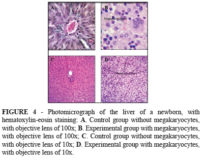
The livers of the control group animals were within the normal pattern for the age group, with the trabecular structure of the hepatocytes maintained. The livers were permeated by rare megakaryocytes and conserved sinusoid capillaries. In the experimental group, morphological-histological hepatic destructuring was observed in all the newborns, with removal of the hepatic trabeculae and severe hepatic extramedullary hematopoiesis, well characterized by severe megakaryocytosis. The following figures illustrate the differences between the experimental and control groups, in relation to the total number of megakaryocytes in the individuals (Figures 1 to 5).
Discussion
With the exposure of populations to pesticides, the consequent possibility that several diseases might be acquired has been raised. Contamination among agricultural workers has been confirmed, with elimination of the drug through the urinary tract11. A significant proportion of the labor force in agriculture is formed by women of fertile age19.
Members of the general population are exposed over the course of their lives to various combinations of pesticides at low doses, mainly through food consumption. However, it is possible that mixtures of pesticides may produce additive, synergic or antagonistic effects5.
Merhi et al.5 demonstrated in France that in rats that received doses that were proportional in body weight terms to the doses received by humans in fruits and vegetables, over a four-week period, the regulatory factors for hematopoiesis became altered. In the present study, exposure of the pregnant female rats led to fetuses with severe musculoskeletal, digestive tract and anorectal abnormalities, as also described by Macedo et al.7 and Lemos et al.20.
Another type of abnormality observed in the fetal digestive system in the present study was hepatic abnormalities, which manifested as distorted morphology, edema and increased volume in 74% of the group exposed to ethylenethiourea. Altered consistency was also observed, with friability in 100% of the newborns. There was also a change in coloration, to wine red, and at the histological level, there were alterations to hematopoietic factors.
The presence of neoplastic lesions in the liver and thyroid, as described in a study conducted by Mattioli et al.9 on adult rats exposed to ethylenethiourea, was not repeated in the fetuses of the present study.
Changes to the production of hematopoietic factors were observed, characterized by foci of myelocytic, erythrocytic and megakaryocytic cells. In the present study, this occurred mainly at the cost of the platelet lineage derived from megakaryoblasts, followed by megakaryocytes, thus portraying severe extramedullary hematopoiesis. This occurs mainly when there is a failure of the medullary hematopoiesis mechanism and the presence of neoplastic diseases.
Hepatic megakaryocytosis in rat fetuses is found physiologically from the 11th day of gestation and increases until the 13th day. Thereafter, it reduces towards the end of the pregnancy. On the eighth day after delivery, only a few megakaryocytes are observed21.
In the present study, after the fetal exposure to ethylenethiourea, the liver was shown to be a site of severe extramedullary hematopoiesis in the animals of the experimental group. The mean megakaryocyte count in these animals was eight times greater than in the newborns in the control group during the same gestational phase of exposure. The mean density of megakaryocytes in the newborns from the mothers in the control group was between 12.5 and 15 megakaryocytes/mm2, while the means for the experimental group ranged from 107.9 to 114.2. This difference between the groups was statistically significant and characterized hepatic extramedullary hematopoiesis among the newborns in the experimental group.
The observed hepatic effects from fetal exposure to ethylenethiourea highlight the importance of exposure to pesticides and its effects relating to hematopoietic dysfunction and consequent hepatic extramedullary hematopoiesis.
Hematopoietic tissues have been found to present higher activity in fetal peripheral blood in cases of severe anemia, with increased erythroblast counts (i.e. erythroblastosis)17. This hematopoiesis causes blood congestion in hepatocytes, which leads to sinusoidal hypertension and consequent hydrops17.
In the present study, 100% of the newborns that were exposed to ethylenethiourea presented hydrops, whereas in the control group, all the newborns were healthy. This finding was also described by Osano et al.16, who also noted that 100% of the newborns exposed to dithiocarbamate and amitraz presented hydrops.
In the newborns with hydrops, alterations to hepatic morphology were observed, and these progressed with persistent severe hepatic extramedullary hematopoiesis beyond the expected gestational period.
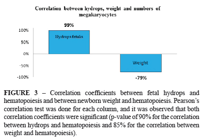
Through correlating the data relating to fetal hydrops, hepatic morphological alterations and hepatic extramedullary hematopoiesis among the newborns in the experimental group, it was found that these presented associations such that 100% of these newborns presented hydrops and hepatic morphological alterations, and they also all presented significantly increased megakaryocyte density, thus also configuring severe extramedullary hematopoiesis in 100% of them. These data again demonstrate the significant risk that the fetuses of from pregnant rats exposed to this drug were subjected to.
A study conducted by Teixeira et al.15 demonstrated an association between anemia and fetal hydrops. Like in a study conducted among human fetuses with hydrops in which an association between hydrops and death was reported, with anemia from non-immune causes as an important causative factor for hydrops22, this association was also found in the present study, in the group of animals exposed to ethylenethiourea. There were also three deaths in the present sample. This study showed that severe hepatic extramedullary hematopoiesis was present in all the newborns exposed to ethylenethiourea; this abnormality has been associated with anemia and has also been attributed to hydrops15,17.
It has been demonstrated that medullary hematopoiesis is stimulated by mild anemia, and that with severe anemia, recruitment of extramedullary sites occurs. Thus, severe hepatic erythropoiesis is thought to cause fetal hydrops17. In this way, an association has been observed between hydrops and the abnormalities of hematopoiesis, anemia or hepatic insufficiency22.
Among the newborns of the experimental group, it was seen that the hydrops presented was associated with significantly delayed fetal growth. The newborns in the control group presented a mean weight of five grams, while those in the experimental group had a mean of 2.5 grams. Thus, the newborns from mothers exposed to ethylenethiourea weighed around half the weight of the newborns in the control group.
A study conducted among pregnant women in Mexico demonstrated that pesticides were present in the umbilical cord, thus showing that these fetuses were contaminated by pesticides23. From observations on the serum levels of contaminants due to pesticides, in blood from the umbilical cord and placental tissue of pregnant women in Canada, such contamination was reported to be an important factor for the presence of delayed fetal growth24.
A study has indicated that frequent and indiscriminate use of pesticides has produced genotoxic damage subsequent to occupational exposure to these substances25. However, few such evaluations have been made among fetuses and newborns: such studies have almost always focuses on adults exposed directly or indirectly. Thus, the consequences for fetuses have not been satisfactorily assessed.
It needs to be emphasized that fetal exposure to pesticides may be an unidentified cause of fetal diseases. Therefore, the present study serves to warn that attention needs to be directed towards evaluating this health problem.
Conclusion
Exposure of pregnant rats to a pesticide derivative gave rise to fetal hepatic damage, with congestion, hepatic friability and morphological-histological hepatic destructuring in all the newborns exposed to ethylenethiourea, with removal of the hepatic trabeculae and severe hepatic megakaryocytosis, thus characterizing extramedullary hematopoiesis. The latter was shown to be an important predisposing factor for hydrops and low fetal weight.
Received: July 19, 2012
Review: September 18, 2012
Accepted: October 17, 2012
Conflict of interest: none
Financial source: none
- 1. Bedor CNG, Ramos LO, Rego MAV, Pavão AC, Augusto LGS. Avaliação e reflexos da comercialização e utilização de agrotóxicos na região do submédio do Vale do São Francisco. Rev Baiana Saúde Pública. 2007;31(1):68-76.
- 2. Soares W, Almeida RMVR, Moro S. Rural work and risk factors associated with pesticide use in Minas Gerais, Brazil. Cad Saúde Pública. 2003;19(4);1117-27.
- 3. Peres F, Moreira JC, Luz C. Os impactos dos agrotóxicos sobre a saúde e o ambiente. Rev Saúde Coletiva. 2007;12(1):4.
- 4. Faria NMX, Fassa AG, Facchini LA. Intoxicação por agrotóxicos no Brasil: os sistemas oficiais de informação e desafios para realização de estudos epidemiológicos. Ciênc Saúde Coletiva. 2007;12(1):25-38.
- 5. Merhi M,Demur C,Racaud-Sultan C,Bertrand J,Canlet C,Estrada FB, Gamet-Payrastre L. Gender-linked haematopoietic and metabolic disturbances induced by a pesticide mixture administered at low dose to mice. Toxicology. 2010;267(1-3):80-90.
- 6. Elzanaty S,Rignell-Hydbom A,Jönsson BA,Pedersen HS,Ludwicki JK,Shevets M,Zvyezday V, Toft G, Bonde JP, Rylander L, Hagmar L, Bonefeld-Jorgensen E, Spano M, Bizzaro D, Manicardi GC, Giwercman A; INUENDO. Association between exposure to persistent organohalogen pollutants and epididymal and accessory sex gland function: multicentre study in Inuit and European populations. Reprod Toxicol. 2006;22(4):765-73.
- 7. Macedo M, Martins JL, Meyer KF. Evaluation of an experimental model for anorectal anomalies induced by ethylenethiourea1. Acta Cir Bras. 2007;22(2):130-6.
- 8. Frakes RA. Drinking water guideline for ethylene thiourea, a metabolite of ethylene bisdithiocarbamate fungicides. Regul Toxicol Pharmacol. 1988;8(2):207-18.
- 9. Mattioli F, Martelli A, Gosmar M, Garbero C, Manfredi V, Varaldo E, Torre GC, Brambilla G. DNA fragmentation and DNA repair synthesis induced in rat and human thyroid cells by chemicals carcinogenic to the rat thyroid. Mutat Res. 2006;609(2):146-53.
- 10. Pandey M, Raizada RB, Dikshith TS. 90-day oral toxicity of ziram: a thyrostatic and hepatotoxic study. Environ Pollut. 1990;65(4):311-22.
- 11. Houeto P, Bindoula G, Hoffman JR. Ethylenebisdithiocarbamates and ethylenethiourea: possible human health hazards. Environ Health Perspect. 1995;103(6):568-73.
- 12. Alegria H, Wong F, Jantunen LM, Bidleman TF, Salvador-Figueroa M, Gold-Bouchot G, Moreno VC, Waliszewski SM, Infanzon R. Organochlorine pesticides and PCBs in air of southern México (20022004). Atmospheric Environ. 2008;42(38):88108.
- 13. Rosada J, Bindi M, Pinelli M, Pandolfo C, Cassetti G, Castiglioni M. Hematopoyesis extramedular: mecanismo compensador o síndrome clínico? Descripción de un caso y revisión bibliográfica. An Med Interna. 2007;24(2):77-80.
- 14. Costa-Val R, Nunes TA, Silva RC, Souza AF, Souza IE, Souza TK. Inhibition of rats extramedullary liver erytropoiesis by hyperbaric oxygen therapy. Acta Cir Bras. 2007;22(2):137-41.
- 15. Teixeira A, Rocha G, Guedes MB, Guimarães H. Recém-nascido com hidrópsia fetal não imune - Experiência de um Centro de Referência. Acta Med Port. 2008;21(4):345-50.
- 16. Osano O, Oladimeji AA, Kraak MH, Admiraal W. Teratogenic effects of amitraz, 2,4-dimethylaniline, and paraquat on developing frog (Xenopus) embryos. Arch Environ Contam Toxicol. 2002;43(1):42-9.
- 17. Nicolaides KH, Thilaganathan B, Rodeck CH, Mibashan RS. Erythroblastosis and reticulocytosis in anemic fetuses. Am J Obstet Gynecol. 1988;159(5):1063-5.
- 18. Alves AC. Histologia da medula óssea. Rev Bras Hematol Hemoter. 2009;31(3):183-8.
- 19. Silva SRG, Martins JL, Seixas S, Silva DCG, Lemos SPP, Lemos PVB. Defeitos congênitos e exposição a agrotóxicos no Vale do São Francisco. Rev Bras Ginecol Obstet. 2011;33(1):20-6.
- 20. Lemos SPP, Martins JL, Lemos PVRB, Silva SRG, Santos FL, Silva Júnior VA. Acta Cir Bras. 2012;27(3):244-50.
- 21. Bockman DE,Gulati AK. Localization of fibronectin in megakaryocytes of fetal liver. Anat Rec.1989;223(1):90-4.
- 22. Léticée N,Bessières-Grattagliano B,Dupré T,Vuillaumier-Barrot S,de Lonlay P,Razavi F, Khartoufi NE, Ville Y, Vekemans M, Bouvier R, Seta N, Attié-Bitach T. Should PMM2-deficiency (CDG Ia) be searched in every case of unexplained hydrops fetalis ? Mol Genet Metab. 2010;101(2-3):253-7.
- 23. Herrero-Mercado M, Waliszewski SM, Caba M, Martínez-Valenzuela C, Hernández-Chalate F. Organochlorine pesticide levels in umbilical cord blood of newborn in Veracruz, Mexico. Bull Environ Contam Toxicol. 2010;85(4):367-71.
- 24. Hamel A, Mergler D, Takser L, Simoneau L, Lafond J. Effects of low concentrations of organochlorine compounds in women on calcium transfer in human placental syncytiotrophoblast. Toxicol Sci. 2003;76:182-9.
- 25. Martínez-Valenzuela C,Gómez-Arroyo S,Villalobos-Pietrini R,Waliszewski S,Calderón-Segura ME,Félix-Gastélum R, Armando Álvarez-Torres. Genotoxic biomonitoring of agricultural workers exposed to pesticides in the north of Sinaloa State, Mexico. Environ Int. 2009;35(8):1155-9.
Publication Dates
-
Publication in this collection
29 Nov 2012 -
Date of issue
Dec 2012
History
-
Received
19 July 2012 -
Accepted
17 Oct 2012 -
Reviewed
18 Sept 2012


