Abstracts
The aim of this study was to compare the resistance of endodontically treated teeth with intraradicular retainer different amounts of remaining coronal structure. Fifty freshly extracted maxillary canines were endodontically treated and randomly assigned to five groups (n=10), as follows: group 1 (control) = teeth with custom cast post and core; group 2 = teeth without remaining coronal structure; group 3, 4 and 5 = teeth with 1 mm, 2 mm and 3 mm of remaining coronal structure, respectively. All specimens in groups 2 to 5 were restored with prefabricated post and resin core. The teeth were embedded in acrylic resin and the fracture strength was measured on a universal testing machine at 45 degrees to the long axis of the tooth until failure. Data were analyzed statistically by one-way analysis of variance and Tukey's test. There was no significant differences (p>0.05) between the control group and group 2, and between groups 3, 4 and 5 (p>0.05). Control group and group 2 had significantly higher resistance strength than groups 3, 4 and 5 (p<0.00001). The findings of this study showed that teeth without remaining coronal structure had significantly higher fracture strength than those with remaining coronal structure (1 mm, 2 mm and 3 mm). When the dental crown was not completely removed, the amount of remaining coronal dentin did not significantly affect the fracture strength of endodontically treated teeth with intraradicular retainer.
posts and core technique; fracture strength; composite resins
O objetivo deste trabalho foi comparar a resistência de dentes tratados endodonticamente restaurados com pinos intra-radiculares apresentando diferentes quantidades de remanescente dentário coronal. Cinqüenta caninos superiores humanos recentemente extraídos foram tratados endodonticamente e divididos aleatoriamente em 5 grupos (n=10), como descrito a seguir: grupo 1 (controle) = dentes com núcleo metálico fundido; grupo 2 = dentes sem remanescente dentário coronal; grupos 3, 4 e 5 = dentes com 1 mm, 2 mm e 3 mm de remanescente dentário coronal, respectivamente. Todos os espécimes dos grupos 2 a 5 foram restaurados com pinos pré-fabricados e resina composta. Os dentes foram embebidos em resina acrílica e a resistência à fratura foi medida em uma máquina de ensaios universal a 45 graus em relação ao longo eixo do dente até que ocorresse a falha. Os dados foram analisados por meio da análise de variância a 1 critério e teste de Tukey. Não houve diferença estatisticamente significante (p>0.05) entre os grupos controle e grupo 2 e entre os grupos 3, 4 e 5. O grupo controle e o grupo 2 apresentaram resultados significantemente maiores que os grupos 3, 4 e 5 (p<0.00001). Os achados deste estudo demonstraram que os dentes sem remanescente dentário coronal tiveram resistência à fratura significantemente maior do que os dentes com remanescente coronal (1 mm, 2 mm e 3 mm). Quando a coroa dental não foi completamente removida, a quantidade de remanescente coronal não afetou significativamente a resistência de dentes tratados endodonticamente com pinos intra-radiculares.
Resistance of teeth with intraradicular retainer
Influence of the remaining coronal structure on the resistance of teeth with intraradicular retainer
Jefferson Ricardo Pereira; Tatiany de Mendonça Neto; Vinícius de Carvalho Porto; Luiz Fernando Pegoraro; Accácio Lins do Valle
Department of Prosthodontics, Faculty of Dentistry of Bauru, University of São Paulo, Bauru, SP, Brazil
Correspondence Correspondence: Dr. Jefferson Ricardo Pereira Rua Antônio Xavier de Mendonça 8-08, apto 5 Vila Tereza, 17012-385 Bauru, SP, Brasil Tel: +55-14-3227-4857 e-mail: jeffripe@rocketmail.com
ABSTRACT
The aim of this study was to compare the resistance of endodontically treated teeth with intraradicular retainer different amounts of remaining coronal structure. Fifty freshly extracted maxillary canines were endodontically treated and randomly assigned to five groups (n=10), as follows: group 1 (control) = teeth with custom cast post and core; group 2 = teeth without remaining coronal structure; group 3, 4 and 5 = teeth with 1 mm, 2 mm and 3 mm of remaining coronal structure, respectively. All specimens in groups 2 to 5 were restored with prefabricated post and resin core. The teeth were embedded in acrylic resin and the fracture strength was measured on a universal testing machine at 45 degrees to the long axis of the tooth until failure. Data were analyzed statistically by one-way analysis of variance and Tukey's test. There was no significant differences (p>0.05) between the control group and group 2, and between groups 3, 4 and 5 (p>0.05). Control group and group 2 had significantly higher resistance strength than groups 3, 4 and 5 (p<0.00001). The findings of this study showed that teeth without remaining coronal structure had significantly higher fracture strength than those with remaining coronal structure (1 mm, 2 mm and 3 mm). When the dental crown was not completely removed, the amount of remaining coronal dentin did not significantly affect the fracture strength of endodontically treated teeth with intraradicular retainer.
Key Words: posts and core technique, fracture strength, composite resins.
RESUMO
O objetivo deste trabalho foi comparar a resistência de dentes tratados endodonticamente restaurados com pinos intra-radiculares apresentando diferentes quantidades de remanescente dentário coronal. Cinqüenta caninos superiores humanos recentemente extraídos foram tratados endodonticamente e divididos aleatoriamente em 5 grupos (n=10), como descrito a seguir: grupo 1 (controle) = dentes com núcleo metálico fundido; grupo 2 = dentes sem remanescente dentário coronal; grupos 3, 4 e 5 = dentes com 1 mm, 2 mm e 3 mm de remanescente dentário coronal, respectivamente. Todos os espécimes dos grupos 2 a 5 foram restaurados com pinos pré-fabricados e resina composta. Os dentes foram embebidos em resina acrílica e a resistência à fratura foi medida em uma máquina de ensaios universal a 45 graus em relação ao longo eixo do dente até que ocorresse a falha. Os dados foram analisados por meio da análise de variância a 1 critério e teste de Tukey. Não houve diferença estatisticamente significante (p>0.05) entre os grupos controle e grupo 2 e entre os grupos 3, 4 e 5. O grupo controle e o grupo 2 apresentaram resultados significantemente maiores que os grupos 3, 4 e 5 (p<0.00001). Os achados deste estudo demonstraram que os dentes sem remanescente dentário coronal tiveram resistência à fratura significantemente maior do que os dentes com remanescente coronal (1 mm, 2 mm e 3 mm). Quando a coroa dental não foi completamente removida, a quantidade de remanescente coronal não afetou significativamente a resistência de dentes tratados endodonticamente com pinos intra-radiculares.
INTRODUCTION
The restoration of endodontically treated teeth is an important aspect of dental practice that involves a range of treatment options of variable complexity. The clinicians are presented with a multifaceted restorative challenge when confronted with endodontically treated teeth complicated by substantial loss of coronal tooth structure and must be able to predict the probability of successfully restoring such teeth.
The likelihood of survival of a pulpless tooth is directly related to the quantity and quality of the remaining dental tissue. A post is commonly placed in an attempt to strengthen the tooth (1,2). However, in vitro and in vivo studies have demonstrated that a dowel does not reinforce endodontically treated teeth, in spite of improving retention when little amount of clinical crown is remaining (3-4).
Although cast post-core restorations are the restorative treatment of choice for endodontically treated teeth, prefabricated post systems have recently become increasingly popular for providing satisfactory results (5-7). Despite efforts to reinforce endodontically treated teeth with internal posts and cores, fractures still occur. Studies (8-9) have shown that roots restored with cast posts exhibited significantly higher forces than prefabricated posts. Notwithstanding the lower resistance, the restorative technique with prefabricated posts and composite resin may be feasible because all fractures occur in the composite resin core, thereby protecting the tooth structure (9). With recent improvements in composite bonding to dentin, actual internal support is now available (3,10-12).
The availability of at least 1.0 mm of coronal tooth structure between the crown preparation shoulder and the tooth-core junction was found to approximately double fracture strength (13). It seems that this extension of coronal tooth structure provides the greatest resistance and retention forms to the crown (14-15). Several authors (4,16-17) have suggested that the tooth should have a minimal amount of 2 mm of coronal structure above the cementoenamel junction (CEJ) to ensure tooth strength. A previous study has not found statistically significant correlation between the amount of remaining coronal structure and fracture resistance, although the findings of other investigation (19) showed that teeth without remaining coronal structure were more resistant than those presenting 2 mm of remaining coronal structure.
The purpose of this study was to compare the fracture strength of teeth with intraradicular retainer and different amounts of remaining coronal structure. Two hypotheses were investigated: 1) teeth with different remaining coronal structure would differ statistically regarding fracture strength; 2) there would be differences between the types of fracture and posts.
MATERIAL AND METHODS
Fifty maxillary canines with similar root sizes recently extracted for periodontal reasons were selected and stored in distilled water. The inclusion criteria were teeth without root carious lesions or fissures and not previously submitted to endodontic treatment.
Each canal was prepared within 1 mm of the radiographic apex. The master apical file was three sizes larger than the initial instrument used. The root canal was instrumented with a conventional step-back technique up to an ISO 35 K-file at the apical constriction, irrigated with 2.5% sodium hypochlorite solution throughout the preparation and dried with paper points. Each canal was obturated by lateral condensation of gutta-percha points against an ISO 35 master gutta-percha cone and Endométhasone (Spécialités Septodont, Saint-Maur. France) root canal sealer. Post preparation was made with a size 5 Largo reamer to remove 9 mm of gutta-percha from the filled canal.
After post space preparation, the teeth were randomly assigned to 5 groups (n=10). In group 1 (control), the coronal portion was removed at the CEJ perpendicular to the long axis. Root canals were restored with a cast post and core by a direct technique in which the post was made with Duralay acrylic resin (Reliance Dental Mfg. Co., Chicago, USA). Copper/aluminum (Cu-Al) alloy (NPGTM, AalbaDent, Cordelio, CA, USA) was used to cast the posts and cores. The cast post-core sets were cemented with ionomer glass cement (Rely X, 3M Dental, Products Division St. Paul, MN, USA) mixed according to the manufacturer's instructions and introduced into each root canal with a lentulo spiral drill (Maillefer Instruments SA, Ballaigues, Switzerland) at low-speed. The post was coated with cement and seated under finger pressure. During cementation, pressure was allowed to release and the post was gently reseated. Excess material was removed and, after curing, the specimens were stored in distilled water.
Teeth in group 2 (Fig. 1) were prepared in the same way as described for the control group, but the canals were restored with prefabricated stainless steel, parallel-sided, serrated posts with tapered end (number 5317, Euro-Post Anthogyr S.A., Sallanches, France). Rely-X cement (3M) was used to cement the prefabricated posts, following the above-mentioned luting procedures. After setting, excess cement was removed with a probe and dentin was etched with 37% phosphoric acid and bonded with Prime & Bond 2.1 (Dentsply Ind. Com, Petropolis, RJ, Brazil) according to the manufacturer's instructions. Cores were fabricated in a standard manner using a core-forming matrix, poly(methyl methacrylate) resin with composite material (Z100; 3M). The composite was placed using the incremental technique.
In groups 3, 4 and 5, the coronal tooth structure was reduced to a flat plane at a height of 1, 2 and 3 mm from the CEJ (proximal, buccal and lingual), respectively, and restored as described in group 2 (Fig. 1).
The teeth were embedded in acrylic resin (Clássico Artigos Odontológicos S/A, São Paulo, SP, Brazil) poured into acrylic resin molds, mounted along their long axis using a surveyor and placed in a cool water bath during resin curing to avoid damage from the exothermic reaction. Each specimen was placed in a custom apparatus (developed by the authors) at 45 degrees to the buccal/lingual axis and subjected to load on a universal testing machine (Dinamometros KRATOS Ltda, São Paulo, SP, Brazil) (Fig. 2). Shear strength was applied at crosshead speed of 0.5 mm/min until fracture. Failure was recorded when there was fracture of the core material with displacement from the post head, or when fracture affected the core or the tooth.
One-way analysis of variance (ANOVA) was used to compare fracture strength means among the five groups. Multiple comparisons by Tukey's test determined which groups were statistically different from the others. The confidence level was 95%.
RESULTS
Fracture strength was recorded when there was root fracture, resin fracture or both, or upon fracture of the remaining coronal structure. Table 1 summarizes the fracture strength means obtained for the 5 groups. ANOVA showed that at least two of the groups differed statistically from the others.Tukey's test did not show significant differences (p>0.05) between the control group and group 2, and between groups 3, 4 and 5 (p>0.05). Control group and group 2 had significantly higher fracture strength than groups 3, 4 and 5 (p<0.00001) (Table 1).
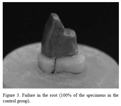
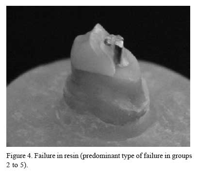
All failures in the control group occurred in the root (Fig. 3). In the other groups, failures predominately occurred in the resin (80% or more) (Table 2, Fig. 4).
DISCUSSION
Restoration by prefabricated posts and composite resin is a viable technique for endodontically treated teeth (5-7). Composite resin fracture occurring when occlusal force is applied may be positive because it may prevent a possible root fracture (9). Root fracture in the cast post-core group (control) and in the group with prefabricated post and no remaining coronal structure (group 2) demanded significantly higher force than that necessary to produce composite resin fracture in the other prefabricated post-core groups (groups 3, 4 and 5), in which remaining coronal structure was present. This fact was related to the higher strength of the nickelchromium alloy, higher modulus of elasticity (8) and high filler content integrated in Z100 resin matrix, possibly due to the size and shape of its particles, which account for 66% of its volume. This higher amount of inorganic load corresponds to the maximum compressive load resistance, surface hardness and wear resistance (3-10). However, under normal clinical conditions the composite resin could resist incisal function. Our results are in agreement with those of a previous study in which composite resin fractured at a lower force than that required to yield root fracture.
Our findings are also consistent with those of other studies (16-17,19), which observed that remaining coronal structure larger than 2 mm was not able to resist compressive load. However, Sorensen (15) showed that 1 mm of remaining coronal structure nearly doubled the fracture resistance. Loney (13) has reported that the maintenance of 1 mm of remaining coronal tooth structure was enough to increase resistance. Isidor (14) agreed with these authors.
In the present study, the remaining coronal structure did not influence the fracture strength of the teeth (Table 2). The results are in agreement with those of Sorensen (4), who maintained 1 and 2 mm of remaining coronal structure and did not observe significant differences between these groups. Conversely, Isidor (14) reported that the increase of remaining coronal structure improved fracture resistance.
The most common cause of failure when direct technique is used (prefabricated post and composite resin) is fracture of the restorative material. This fact was observed in this study and is in agreement with the results of a previous study (9).
It was demonstrated that roots restored by individual cast posts exhibited significantly higher fracture forces than those restored by prefabricated post and resin core. Despite its lower resistance, restoration with prefabricated posts and composite resin may be feasible because there is no root fracture, and thus this technique protects the tooth structure (9). Furthermore, the forces responsible for failure in this study were considerably higher than the maximal physiologic forces acting on the teeth in the oral cavity. Lyons (20) showed that the maximum force applied on the canine is 22 Kgf. In the presence of parafunction, this force is raised to 26 Kgf, and the maximum forces used by this author were between 35 and 37 kgf.
The results of this study showed that there were no significant differences among the groups with remaining coronal structure. Teeth without remaining coronal structure had significantly higher fracture strength than those with remaining coronal structure (1 mm, 2 mm and 3 mm). The control group presented higher fracture strength than the other groups. The predominant mode of failure in the control group showed the worst prognosis because it led to root fracture. On the other hand, the groups restored with prefabricated post and resin core failed in the resin, without damaging the root, thereby protecting the tooth structure.
Accepted June 27, 2005
- 1. Assif D, Gorfil C. Biomechanical considerations in restoring endodontically treated teeth. J Prosthet Dent 1994;71:565-567.
- 2. Cohen BI, Pagnillo M, Condos S, Deutsch AS. Comparison of torsional forces at failure for seven endodontic post systems. J Prosthet Dent 1996;74:350-357.
- 3. Guzy GE, Nicholls JI. In vitro comparison of intact endodontically treated teeth with and without endo-post reinforcement. J Prosthet Dent 1979;42:39-44.
- 4. Sorensen JA, Engelman MJ. Ferrule design and fracture resistance of endodontically treated teeth. J Prosthet Dent 1990;63:529-536.
- 5. Hopwood WA, Wilson NH. Clinical assessment of split-shank post system in permanent molar and pre-molar teeth. Quintessence Int 1990;21:907-911.
- 6. Stockton LW. Factors affecting retention of post systems: a literature review. J Prosthet Dent 1999;81:380-385.
- 7. Torbjorner A, Karlsson S, Odman PA. Survival rate and failure characteristics for two post designs. J Prosthet Dent 1995;73:439-444.
- 8. Assif D, Oren E, Marshak BL, Aviv I. Photoelastic analysis of stress transfer by endodontically treated teeth to the supporting structure using different restorative techniques. J Prosthet Dent 1989;61:676-678.
- 9. Fraga RC, Chaves BT, Mello GS, Siqueira JF Jr. Fracture resistance of endodontically treated roots after restoration. J Oral Rehab 1998;25:809-813.
- 10. Abdalla AI, Alhadainy HA. 2-year clinical evaluation of class I posterior composites. Am J Dent 1996;9:150-152.
- 11. Bex RT, Parker MW, Judkins JT, Pelleu GB Jr. Effect of dentinal bonded resin post-core preparations on resistance to vertical fracture. J Prosthet Dent 1992;67:768-772.
- 12. Bowen RL, Cobb EN. A method for bonding to dentin and enamel. J Am Dent Ass 1983;107:1070-1076.
- 13. Loney RW, Kotowics WE, Mcdowell GC. Three-dimension photoelastic stress analysis of the ferrule effect in cast post and cores. J Prosthet Dent 1990;63:506-546.
- 14. Isidor F, Brondum K, Ravnholt G. The influence of post length and crown ferrule on the resistance to cyclic loading of bovine teeth prefabricated titanium post. Int J Prosthodont 1999;12:79-82.
- 15. Sorensen JA. Preservation of tooth structure. Can Dent Ass J 1988;5:15-21.
- 16. Goodacre CJ. Five factors to be considered when restoring treated teeth. Pract Proced Aesthet Dent 2004;16:455-460.
- 17. Morgano SM, Rodrigues AHC, Sabrosa CE. Resoration of endodontically treated teeth. Dent Clin N Amer, 2004;48:397-416.
- 18. Gegauff AG. Effect of crown lengthening and ferrule placement on static load failure of cemented cast post-cores and crowns. J Prosthet Dent 2000;84:169-179.
- 19. Sorensen JA, Engelman MJ. Effect of post adaptation on fracture resistance of endodontically treated teeth. J Prosthet Dent 1990;64:419-424.
- 20. Lyons MF. A preliminary electromyographic study of bite force and jaw-closing muscle fatigue in human subjects with advanced tooth wear. J Oral Rehab 1990;17:311-318.
Publication Dates
-
Publication in this collection
12 Jan 2006 -
Date of issue
Dec 2005
History
-
Accepted
27 June 2005 -
Received
27 June 2005


