Abstracts
The authors review 12 studies about posterior cruciate ligament isometry. The authors conclude that most studies agree that femoral insertion of the ligament is more important for isometry, and that there is a most isometric line or area inside the femoral insertion, perpendicular to the roof of the intercondilar fossa, approximately 10 to 14mm from its anterior opening.
Biomechanics; Injuries; Injuries
Trabalho de revisão bibliográfica referente à isometricidade do ligamento cruzado posterior. São avaliados doze artigos que estudam a isometricidade do ligamento, constatando que a maioria destes é concorde com a maior importância da inserção femoral na isometricidade e que existe uma linha ou área mais isométrica na inserção femoral, aproximadamente perpendicular ao teto da fossa intercondilar, localizada de 10 a 14mm da abertura anterior desta fossa.
Biomecânica; Lesões; Lesões
ARTIGO DE REVISÃO
Isometry of the posterior cruciate ligament
Alexandre E. V. KokronI; Arnaldo J. HernandezII; Gilberto L. CamanhoIII; Marco M. AmatuzziIV
IDoutor em Ortopedia
IIDoutor do IOT - HC - FMUSP
IIIProfessor Livre Docente do IOT
IV Professor Titular e Chefe do Departamento de Ortopedia e Traumatologia da Faculdade de Medicina da Universidade de São Paulo
SUMMARY
The authors review 12 studies about posterior cruciate ligament isometry. The authors conclude that most studies agree that femoral insertion of the ligament is more important for isometry, and that there is a most isometric line or area inside the femoral insertion, perpendicular to the roof of the intercondilar fossa, approximately 10 to 14mm from its anterior opening.
Key words: Biomechanics - Posterior Cruciate Ligament/ Injuries - Posterior Cruciate Ligament/ Surgery
INTRODUCTION
To reconstruct the posterior cruciate ligament (LCP) it is fundamental to know its isometry. The LCP isometry was studied by several authors using different experimental models, as unextensible wire connecting the points or trigonometric calculus, (Bach et al.3, Bomberg et al.4, Covey et al.5, Friederich and O'Brien6, Grood et al.8, Juergensen et al.9, Kurosawa et al.11, Odensten and Gillquist12, Ogata and McCarthy13, Ortiz et al.14, Sidles et al.15, Trus et al.16).
The aim of this study was to summarize information from the literature as concerns isometry of the posterior cruciate ligament.
The literature is confuse as concerns guiding directions inside the intercondylar fossa, since two nomenclatures are used: the anatomical, which considers directions with the extended knee, and the surgical, based on a 90 degrees bent knee (Amis et al.1). To avoid this, the European Society of Sports Medicine, Knee Surgery and Arthroscopy suggests that the anatomical terminology is used concerning positioning inside the intercondylar fossa (Amis et al.1). Considering that in the sagittal plane the roof of the intercondylar fossa is oblique in relation to the anterior-posterior and proximal-distal directions, if along this roof a point is anterior to another it is automatically distal to the latter; however, when a point is said to be in a more posterior position along the roof of the intercondylar fossa, that point is also more proximal (Amis et al.1).
Results from studies which compare variation in isometry caused by modification in the localization of femur and tibia points show uniformity since modifications in the tibia cause small alterations in isometry, while changes in the localization of points in the femur can cause large variations in isometry (Bach et al.3, Bomberg et al.4, Friederich and O'Brien6, Grood et al.8, Juergensen et al.9, Sidles et al.15, Trus et al.16).
There are controversies in the literature concerning the PCL most isometric area in the femur. Most of the studies observe that modifications along the roof of the intercondylar fossa to the anterior (distal) or posterior (proximal) positions cause important variations in the fibers isometry and also that modifications in a direction perpendicular to the roof cause small variations in isometry (Bomberg et al.4, Friederich and O'Brien6, Grood et al.8, Ogata and McCarthy13, Sidles et al.15, Trus et al.16). Accordingly, inside the anatomical insertion of the PCL some authors define an isometric line or area, approximately perpendicular to the roof of the fossa, dividing its insertion in two non-isometric areas; the isometric area is about 3 mm wide, and the inserted fibers present an isometric or almost isometric behavior (Friederich and O'Brien6, Grood et al.8, Sidles et al.15, Trus et al.18). Most of the PCL fibers are inserted in the distal (anterior) non-isometric area and are lengthened 4 to 8 mm with a bent knee, loosening in extension; the fibers inserted in the proximal (posterior) non-isometric area lenghten in extension and loose in flexure (Back et al.3, Bomberg et al.4, Covey et al.5, Friederich and O'Brien6, Grood et al.8, Juergensen et al.9, Kurosawa et al.11, Odensten and Gillquist12, Ogata and McCarthy13, Sidles et al.15, Trus et al.16).
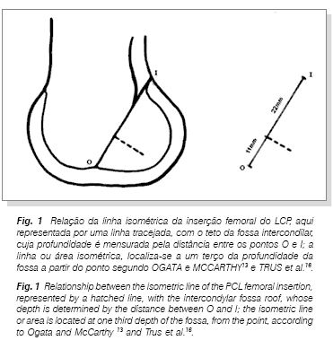
Some authors describe the localization of this isometric line in the PCL femoral insertion as the distance in milimeters along the intercondylar fossa roof until the cartilage-bone junction in the anterior opening of the fossa, while others prefer to use the third of the roof depth rule or use the ligament insertion as reference (Back et al. 3, Bomberg et al.4, Covey et al.5, Friederich and O'Brien6, Grood et al.8, Juergensen et al.9, Odensten and Gillquist12, Ogata and McCarthy13. Ortiz et al.14, Sidles et al.15, Trus et al.16). When expressed in absolute numbers, the isometric line is located along the intercondylar fossa roof at 13 mm according to Sidles et al.15 and 11 mm according to Grood et al.8. The third of the depth rule, used by Ogata and McCarthy13 and by Trus et al.16, consists of measuring the distance between the anterior and posterior opening of the intercondylar fossa, and dividing it by three; the result of this division is the isometric line depth from the cartilage-bone junction of the anterior opening (figure 1). According to Ogata and McCarthy13 and Trus et al.16, the results were 10 mm and 11 mm, respectively. In a national study, Kokron10 observes that the intercondylar fossa depth is 31.6 mm averaging 14 knees (minimum 29 mm and maximum 34 mm). Thus, one third of these numbers corresponds to 10 or 11 mm, as those described by Ogata and McCarthy13 and Trus et al.16.
Other authors prefer to use as reference the femoral anatomical insertion concerning the site of the isometric point, as described by Covey et al.5 and Friederich and O'Brien6. We do not consider this reference adequate for knees with chronic ligament lesion when this parameter can show alterations.
The isometric area of the PCL femoral insertion can vary due to several factors: direction of the force applied to the tibia, presence or absence of articular spacing due to weight of the leg or foot, presence of muscular contraction, and even integrity or lack of integrity of the PCL (Covey et al.5, Grood et al.8, Kurosawa et al.11, Ortiz et al.14). The influence of muscular contraction on the PCL isometry was initially reported by Kurosawa et al.11 in 1991, who observed that the application of a force on the patella simulating contraction of the quadriceps lessens the lenght of the PCL fibers in several angles of knee flexure as compared to the evaluation without the application of force. In the Ortiz et al.14 study, simulating in laboratory situations of muscular contraction corresponding to daily life activities, the localization of the PCL isometric point is so much modified that it remains outside the ligament femoral insertion. The very absence of the PCL produces alterations in isometry, since the absence of primary restriction on the posterior displacement of the tibia allows it to stay in a more posterior position during all the arch of movement of the knee, similarly to what occurs with isometry when the ACL is absent (Amis and Jakob2, Covey et al.5, Furia et al.7).
We prefer to locate the femur isometric line using the rule of the third, since it considers knee size; we are aware that the results using this value are consistent with Grood et al.8's results. The localization of the center in the tibia tunnel somewhat alters the graft isometry, since it is inside the ligament anatomical insertion.
The controversy concerning the use of the femoral isometric tunnel or the femoral non-isometric tunnel (anterior to the isometric line) will be described in another review.
REFERENCES
Orthopedics and Traumatology Institute, "Hospital das Clínicas", University of São Paulo College of Medicine
- 1. AMIS, A.A., BEYNNON, B., BLANKEVOORT, L., CHAMBAT, P., CHRISTEL, P., DURSELEN, L., FRIEDERICH N., GROOD, E., HERTEL, P., JAKOB, R.P., MULLER, W., O'BRIEN, M. & O'CONNOR, J.: Proceedings of the ESSKA scientific workshop on reconstruction of the anterior and posterior cruciate ligaments. Knee Surg. Sports Traumatol. Arthrosc., 2:124-32 , 1994.
- 2. AMIS, A.A. & JAKOB, R.P.: Anterior cruciate ligament graft positioning, tensioning and twisting. Knee Surg. Sports Traumatol. Arthrosc., 6:2-12, 1998. Supplement 1.
- 3. BACH B.R., DALUGA, D.J., MIKOSZ, R., ANDRIACCHI, T.P. & SEIDL, R.: Force displacement characteristics of the posterior cruciate ligament. Am. J. Sports Med., 20: 67-72, 1992.
- 4. BOMBERG, B.C., ACKER, J.H., BOYLE, J. & ZARINS, B.: The effect of posterior ligament loss and reconstruction on the knee. Am. J. Knee Surg., 3:85-96, 1990.
- 5. COVEY, D.C., SAPEGA, A.A. & SHERMAN, G.M.: Testing for isometry during reconstruction of the posterior cruciate ligament: anatomic and biomechanical considerations. Am. J. Sports Med., 24:740-6, 1996.
- 6. FRIEDERICH, N.F. & O'BRIEN, W.R.: Functional anatomy of the cruciate ligaments; in: Jakob, R.P. & Stäubli H.-U., ed. The knee and the cruciate ligaments. Berlin, Springer-Verlag, 1992. p. 78-91
- 7. FURIA, J.P., LINTNER, D.M., SAIZ, P., KOHL, H.W. & NOBLE, P.: Isometry measurements in the knee with the anterior cruciate ligament intact, sectioned, and reconstructed. Am. J. Sports Med, 25:346-52, 1997.
- 8. GROOD, E.S., HEFZY, M.S. & LINDENFIELD, T.N.: Factors affecting the region of most isometric femoral attachments. Am. J. Sports Med., 17: 197-207, 1989.
- 9. JUERGENSEN, K., EDWARDS, J.C. & JAKOB, R.P.: Positioning of the posterior cruciate ligament. Knee Surg. Sports Traumatol. Arthrosc., 2:133-7, 1994.
- 10. KOKRON, A.E.V.: Avaliação da estabilidade da reconstrução na lesão isolada do ligamento cruzado posterior com um e dois feixes de enxerto. Estudo experimental com tendão do músculo quadríceps da coxa e com tendões dos músculos semitendíneo e grácil. São Paulo, 2000. Tese (Doutorado) _ Faculdade de Medicina, Universidade de São Paulo.
- 11. KUROSAWA, H., YAMAKOSHI, K-I., YASUDA, K. & SASAKI, T.: Simultaneous measurement of changes in length of the cruciate ligaments during knee motion. Clin. Orthop., 265:233-9, 1991.
- 12. ODENSTEN, M. & GILLQUIST, J.: Reconstruction of the posterior cruciate ligament using a new drill-guide. Knee Surg. Sports Traumatol. Arthrosc., 1, 39-43, 1993.
- 13. OGATA, K. & MCCARTHY, J.Á.: Measurements of length and tension patterns during reconstruction of the posterior cruciate ligament. Am. J. Sports Med., 20:351-5, 1992.
- 14. ORTIZ, G.J., SCHMOTZER, H., BERNBECK, J., GRAHAM, S., TIBONE, J.E. & VANGSNESS, C.T., JR.: Isometry of the posterior cruciate ligament. Am. J. Sports Med., 26: 663-8, 1998.
- 15. SIDLES, J.A., LARSON, R.V. & GARBINI, J.L.: Ligament length relationships the moving knee. J. Orthop Res., 6:593-610, 1988.
- 16. TRUS, P., PETERMANN, J. & GOTZEN, L.: Posterior cruciate ligament (PCL) reconstruction - an in vitro study of isometry. Part I. Tests using a string linkage model. Knee Surg. Sports Traumatol. Arthrosc., 2:100-3, 1994.
Publication Dates
-
Publication in this collection
17 May 2006 -
Date of issue
June 2001