 Hydroxyapatite-lignin composite as a metallic implant-bone tissue osseointegration improver: experimental study in dogs
Abstract
The study aimed to evaluate biocompatibility, osteoconduction and osseointegration of a pasty composite of hydroxyapatite (20%) and lignin (80%) as a promoter of metal implant and bone tissue integration. An intramedullary Schanz pin was implanted in both tibias of fifteen bitches. In the left tibia, the pin was coated with the biomaterial at the time of surgery. Marrow cavity was also filled with the biomaterial. Right limb did not receive the biomaterial, then constituting the control group. Tibias were harvested from five animals at 8, 60 and 150 days after surgery; three of them were analyzed by histological and biomechanical assessment and the two remaining tibias by X-ray diffraction. Results showed that the biomaterial is biocompatible, with osteoconductivity and osseointegration properties. Histological analysis and diffractograms showed the presence of hydroxyapatite in samples in all periods, although the presence of organic material of low crystallinity was variable. There was no statistical difference in the forces required for removal of the biocompatibility, osteoconductivity and osseointegration, it was not able to promote a better intramedullary pin anchorage.
Key words:
biomaterial; biocompatibility; osseointegration; osteoconduction; tissue regeneration
Resumo
O trabalho objetivou avaliar a biocompatibilidade, a osteocondução e a osseointegração de um compósito pastoso de hidroxiapatita (20%) e lignina (80%) como promotor de integração entre implante metálico e tecido ósseo. Um pino intramedular de Schanz foi implantado em ambas as tíbias de quinze cadelas. Na tíbia esquerda, o pino foi coberto com o biomaterial no momento da cirurgia. A cavidade medular também foi preenchida com o biomaterial. O membro direito não recebeu o biomaterial, constituindo assim o grupo controle. As tíbias foram coletadas de cinco animais aos oito, 60 e 150 dias após a cirurgia, sendo três encaminhadas para avaliação histológica e biomecânica e as duas restantes, para difração de raios X. Os resultados revelaram biocompatibilidade do material, com propriedades de osteocondução e osseointegração. As análises histológicas e os difratogramas mostraram presença da hidroxiapatita no tecido em todos os períodos avaliados, embora a presença de material orgânico de baixa cristalinidade tenha sido variável. Não houve diferença estatística na força necessária para remoção do pino intramedular entre os tempos ou tratamentos. Embora o biomaterial tenha apresentado biocompatibilidade, osteocondutividade e osseointegração, ele não foi capaz de promover uma melhor ancoragem do pino intramedular.
Palavras-chave:
biomaterial; biocompatibilidade; osseointegração; osteocondução; regeneração tecidual
INTRODUCTION:
Metallic implants are widely used in a variety of orthopedic and dental procedures, both in medicine and in veterinary medicine. Erosion of implant or cement used to attach the implant to the bone can produce debris that induce osteolysis triggered by inflammatory response, leading to implant loosening (JIANG et al., 2013). Coating the implants has been suggested in order to avoid this problem (EPINETTE & MANELY, 2007). Studies using hydroxyapatite as biomaterial in bone repair have proved its osteoconductive and biocompatibility (CARLO et al., 2009; FABBRI et al., 2010); therefore, its use as bone cement can be useful. These same authors have commented that hydroxyapatite associated with polymers can enhance its properties. Lignin is a complex natural polymer that is biocompatible and has great potential in association with hydroxyapatite as a biomaterial for bone repair (MARTÍNEZ et al., 2009a) and metal implants coating (ERAKOVIĆ et al., 2012). Moreover, injectable biomaterial containing hydroxyapatite may be effective in regenerating defects adjacent to a metallic implant (CRESPI et al., 2012). Thus, combination of hydroxyapatite and lignin is expected to work as a resorbable bone cement promoting metallic implant osseointegration while supports bone regeneration, lowering the risks of implant loosening. The use of prostheses are growing in veterinary medicine, especially in rehabilitation of small animals, and loosening of metallic implants are important complications in dogs (HETTLICH et al., 2013). Therefore, the aim of the present study was to evaluate biocompatibility, osteoconductivity and osseointegration of a composite made of 20% hydroxyapatite and 80% lignin and its role in bone formation and anchoring of an intramedullary pin in dogs' tibia The presentation developed for this study would prevent the described problem and would allow to obtain biomaterial of low cost, thus more accessible.
MATERIAL AND METHODS:
A composite combining synthetic hydroxyapatite (20%) and lignin (80%) was produced as solid chalky tablets weighing 200mg (HAP-91(r), JHS Biomateriais, Brazil). Fifteen mongrel spayed bitches, aged 1-2 years old, skeletally mature at radiographic examination, and weighing 10-15kg were used in this study. They were kept in individual boxes and food was given twice per day and water available ad libitum . With 12-hour of water and solid fastening, animals received cefazolin (20mg kg-1, intravenous) (Cefazolina sódica, Aurobindo, Brazil) 30 minutes prior to anesthesia induction. They were then sedated with acepromazine (0.1mg kg-1, intravenous) (Acepran(r) 1%, Univet, Brazil) and general anesthesia was induced with propofol (6mg kg-1, intravenous) (Propofol(r), Eurofarma, Brazil) and maintained with isoflurane diluted in 100% O2 (inhalation) (Isofluorano(r), Cristália, Brazil). For a better intraoperative analgesia, epidural anesthesia was performed using morphine (0.1mg kg-1) (Dimorf(r), Cristália, Brazil) combined with lidocaine (0.2mL kg-1) (Xylestesin(r) 2%, Cristália, Brazil). Then a cortical bone defect was created in the proximal diaphysis of the right tibia. The defect was created using an electric drill (Makita, Pet Medical, Brazil) with 4.5mm in diameter and depth enough to reach medullary canal. An intramedullary Steinnamn pin was repeatedly introduced and removed through the defect to detach bone marrow that was partly removed with a 14G catheter and a syringe. Approximately 3mL of this bone marrow was mixed with one tablet of the composite (200mg) and then introduced in the medullary canal. The threaded end of an intramedullary Schanz pin (alloy F138, Ortovet, Brazil) was dipped into the resultant paste of composite and bone marrow and then it was inserted into the medullary canal. The remaining paste was introduced in the canal with a 14G catheter and a syringe, in order to completely fill the defect. The Schanz pin was chosen for each animal based on pre-operative radiographs so it would fill approximately 80% of the most distal part of the canal, ranging from 2.0 to 3.0.Left limb was the control treatment, in which the same surgical procedures were performed, but without the composite. Postoperatively, animals received tramadol (6mg kg-1, for the first 24 hours) (Tramadol(r), União Química, Brazil) and carprofen (4.4mg kg-1, once a day, for three days) (Carproflan(r), Agener União, Brazil) and had surgical wound cleaned daily until complete healing. In the 8th, 60th and 150th days after surgery, five animals randomly underwent euthanasia by overdose of thiopental (20mg kg-1, intravenous) (Tiopental(r) 1g, Cristália, Brazil) and cardiac arrest with potassium chloride 10% (~30mL per animal, intravenous) (Cloreto de Potássio 10%, Isofarma Industrial Farmacêutica, Brazil). Both tibias from three animals were evaluated by biomechanical and histological analysis and those from the remaining two animals were submitted to X-ray diffraction (XRD) analysis.
Biomechanical evaluation was performed in a universal tensile test machine Instron(r). Proximal diaphysis was removed to expose the pin that was attached to claws in which a tension force of 0.5g was applied with a constant displacement rate of 1mm min-1. Tensile strength was obtained in kgf. A proximal fragment containing the defect region was taken for histological analysis. Samples were fixed in 10% buffered formalin, decalcified in formic acid and sodium citrate, dehydrated in alcohol, cleared in xylene and embedded in paraffin and 5-mm sections were stained by hematoxylin and eosin. Types of the tissue and tissue/biomaterial interface features in the defect region were analyzed under light microscopy (Olympus CX31) and described. For crystallinity analysis, bones were dried in an electric oven at 70ºC for 72 hours and then bone marrow was scraped. In addition to this preparation, the composite alone and pure synthetic hydroxyapatite were analyzed in a PANalytical's X'Pert PRO diffractometer. Tensile forces were analyzed by Kruskal-Wallis test for date comparison and by paired t-test for treatment comparison. The tests were conducted in SAS 9.2 software considering a 5% significance level. Histological and crystallinity analyses are presented by descriptive evaluation.
RESULTS AND DISCUSSION:
The composite consistency acquired allowed easy handling and application, but it showed low adhesion and could be washed out by bleeding at the surgical site. RIGO et al. (2007) found similar problems when using particulate hydroxyapatite leading to dispersion and loss of particles. Unlike other studies (BORGES et al., 2000; DUARTE et al., 2006), hydroxyapatite did not provoke more pronounced bleeding than in control animals, probably due to its lower concentration. There were no signs of infection or dehiscence on the surgical wound during the postoperative period.
Biomechanical test recorded maximum resistance provided by newly formed tissues to keep the pin attached to the site where it was implanted. Absolute values increased considerably over time but statistical analysis could not detect significant differences between time (P=0.079 and P=0.061, for treated and control tibia, respectively) and treatments (P=0.655) (Table 1). That may be due to small number of samples and the high variance between the observations. MARTÍNEZ et al. (2009b) obtained similar results when using a composite of hydroxyapatite with 1% of lignin associated with metallic implant in rabbits. In the present study, the defect was filled by provisional matrix containing bone matrix lined by active osteoblasts in both treatments at the 8th day after surgery (Figures 1A and 1B). That was better marked in the treated limb, which may be related to the presence of hydroxyapatite, which has the ability to accelerate bone growth in the first week of bone repair (DAMIEN et al., 2003). No difference between treatments could be noticed at 60th and 150th days after surgery, when there was still new bone, indicating that the remodeling process remained active. There was no formation of fibrous capsule and inflammatory infiltration related to the biomaterial at any moment, suggesting biocompatibility. Although in lesser amounts, the biomaterial was still present at the day 150, but there was no evidence of phagocytosis in histological sections, as previously described by BORGES et al. (2000) and CARLO et al. (2009). ALVES et al. (2011), who applied hydroxyapatite-polyhydroxybutyrate composite in cats, found fibrous capsule formation in response to a chronic inflammatory process, suggesting that this type of reaction could generate mechanical failure of the implant. In this study, the composite presented direct contact with bone matrix, without interposition of other type of tissue, indicating osseointegration (Figure 1C). Acidophilic, acellular and amorphous material, interspersed with small voids, which may correspond to areas where there were hydroxyapatite crystals that were lost in decalcification. BORGES et al. (2000) described histologically hydroxyapatite as amorphous material, acellular and well defined, without affinity by hematoxylin and eosin.

Photomicrograph of the region where the defect for intramedullary pins insertion was created. Treated (A) and control (B) limbs after 8 days: Original bone (a), new bone (b), provisional matrix (c), osteoclasts (black arrow head), biomaterial (white arrow head). Treated limb after 60 days (C): Trabecular bone with osteocytes (a), bone marrow cells (b), biomaterial remains (c), interaction between bone tissue and biomaterial (black arrows). The bar corresponds to 50μm. H&E.
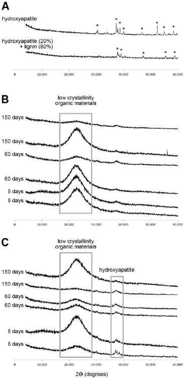
The XRD patterns of hydroxyapatite and hydroxyapatite-lignin composite showed similar peaks (Figure 2A), which is characteristic of ceramics, as described by YAMADA et al. (2012). XRD cannot detect lignin because there is no mineral component in it. A typical wave for organic materials was observed in control treatment but just one peak in one 150th-day sample could be detected (Figure 2B). In one sample from 60th day and another one from 150th day, there was no presence of that wave suggesting that less tissue formation occurred in these animals (Figure 2C). Controlled degradability, namely the gradual replacement of biomaterial components by body's tissues is a desirable feature to promote tissue regeneration (SHOKROLLAHI et al., 2014). Moreover, materials containing calcium phosphate, such as hydroxyapatite, induce osteoblastic differentiation, which also contributes to the osseointegration (RAJZER et al., 2014). Thus, results from XRD and histological analyses, indicate that the composite may have a low rate degradability associated with increased activation and proliferation of osteoblasts, contributing to osseointegration, but not enough to promote satisfactory metal implant anchoring.

X-ray diffraction (XRD) patterns of (A) pure synthetic hydroxyapatite and hydroxyapatite 20% and lignin 80% composite, (B) samples from control and (C) treated limbs 8, 60 and 150 days after application. Waves seen in B and C and absent in A indicates low crystallinity organic materials, which is variable in each sample. Peaks seen in A set hydroxyapatite crystallinity.
According to WAANDERS et al. (2010), viscosity of the bone cement is directly proportional to its penetration into the bone and this is an important feature affecting mechanical cement-bone stability for immediate and long-term implant feasibility. In this study, lignin was used in order to favor intramedullary pin fixation while bone formation promoted by hydroxyapatite occurs. However, the composite obtained after mixing with bone marrow showed spongy consistency and low adherence. On a more applied usage, HUNT et al. (2011), working with methylmethacrylate in hip arthroplasty, also stated that the immediate characteristics of bone-cement-implant interface is one of the most important factors that contribute to initial mechanical fixation and consequent improvement of clinical outcome. One strategy is coating metal implants with hydroxyapatite that produces stable fixation in hybrid prostheses in arthroplasty (PEREIRA et al., 2007). Moreover, the attachment promoted by hydroxyapatite is durable due to bone formation where the ceramic is absorbed (TONINO et al., 2009). Unlike hydroxyapatite-coated implants used in arthroplasty, the present study proposes a biomaterial that can fill the spaces between bone and implant at the moment of the surgery. Lignin is biocompatible and has antioxidant activity (UGARTONDO et al., 2008). It is used in association with hydroxyapatite to cover metal implants such as hip prosthesis in a process that includes sintering, but this procedure may confer cytotoxicity to the biomaterial, when the concentration of lignin is high (ERAKOVIĆ et al., 2012). Additionally, the composite used in the present study is mixed with bone marrow, which brings osteoinduction properties to the biomaterial, a property showed by DEN BOER et al. (2003).
CONCLUSION:
The composite made of 80% of lignin and 20% of hydroxyapatite is biocompatible and has osteoconductivity and osseointegration properties; however, it is not enough to promote better anchorage of the intramedullary pin to bone tissue in the tibia of dogs
ACKNOWLEDGEMENTS
The authors thank Conselho Nacional de Desenvolvimento Científico e Tecnológico (CNPq), Coordenação de Aperfeiçoamento de Pessoal de Nível Superior (CAPES) and Fundação de Amparo à Pesquisa do estado de Minas Gerais (FAPEMIG) for financial support, JHS Ltda. for providing the composite and Prof. Mauricio Fontes (UFV) for XRD analyses.
References
-
ALVES, E.G. et al. Orthopedic implant of a polyhydroxybutyrate (PHB) and hydroxyapatite composite in cats. Journal of Feline Medicine and Surgery, v.13, p.546-552, 2011. Available from: <Available from: <http://jfm.sagepub.com/content/13/8/546.long
>. Accessed: Aug. 06, 2015. doi: 10.1016/j.jfms.2011.03.002.
» https://doi.org/10.1016/j.jfms.2011.03.002» http://jfm.sagepub.com/content/13/8/546.long -
BORGES, A.P.B. et al. Sinthetic hydroxyapatite as bone substitute in the experimentally caused defect on the dog tibia proximal third: Electronic transmission microscopy aspects. Arquivo Brasileiro de Medicina Veterinária e Zootecnia, v.52, n.6, p.616-620, 2000. Available from: <Available from: <http://www.scielo.br/scielo.php?script=sci_arttext&pid=S0102-09352000000600011
>. Accessed: Aug. 06, 2015. doi: 10.1590/S0102-09352000000600011.
» https://doi.org/10.1590/S0102-09352000000600011» http://www.scielo.br/scielo.php?script=sci_arttext&pid=S0102-09352000000600011 -
CARLO, E.C. et al. Tissue response to the composite 50% hydroxyapatite: 50% polyhydroxybutyrate for bone substitution in rabbits. Arquivo Brasileiro de Medicina Veterinária e Zootecnia, v.61, n.4, p.844-852, 2009. Available from: <Available from: <http://www.scielo.br/scielo.php?script=sci_arttext&pid=S0102-09352009000400011&lng=en&nrm=iso&tlng=pt
>. Accessed: Aug. 08, 2015. doi: 10.1590/S0102-09352009000400011.
» https://doi.org/10.1590/S0102-09352009000400011» http://www.scielo.br/scielo.php?script=sci_arttext&pid=S0102-09352009000400011&lng=en&nrm=iso&tlng=pt -
CRESPI, R. et al. Injectable magnesium-enriched hydroxyapatite putty in peri-implant defects: A histomorphometric analysis in pigs. International Journal of Oral & Maxillofacial Implantsv.27, n.1, p.95-101, 2012. Available from: <Available from: <http://www.robertocrespi.it/wp-content/uploads/2012/09/CRESPI-2012-IJOMI.pdf
>. Accessed: Feb. 26, 2014.
» http://www.robertocrespi.it/wp-content/uploads/2012/09/CRESPI-2012-IJOMI.pdf -
DAMIEN, E. et al. A preliminary study on the enhancement of the osteointegration of a novel synthetic hydroxyapatite scaffold in vivo. Journal of Biomedical Materials Research Part A, v.66, n.2, p.241-246, 2003. Available from: <Available from: <http://onlinelibrary.wiley.com/doi/10.1002/jbm.a.10564/full
>. Accessed: Aug. 06, 2015. doi: 10.1002/jbm.a.10564.
» https://doi.org/10.1002/jbm.a.10564» http://onlinelibrary.wiley.com/doi/10.1002/jbm.a.10564/full -
DEN BOER, F.C. et al. Healing of segmental bone defects with granular porous hydroxyapatite augment with recombinant human osteogenic protein-1 or autologous bone marrow. Journal of Orthopaedic Research, v.21, p.521-528, 2003. Available from: <Available from: <http://onlinelibrary.wiley.com/doi/10.1016/S0736-0266(02)00205-X/epdf
>. Accessed: Aug. 06, 2015. doi: 10.1016/S0736-0266(02)00205-X.
» https://doi.org/10.1016/S0736-0266(02)00205-X» http://onlinelibrary.wiley.com/doi/10.1016/S0736-0266(02)00205-X/epdf -
DUARTE, T.S. et al. Osteointegration of synthetic hydroxyapatite in alveolar process of mandible in dogs: histological aspects. Arquivo Brasileiro de Medicina Veterinária e Zootecnia, v.58, n.5, p.848-853, 2006. Available from: <Available from: <http://www.scielo.br/scielo.php?script=sci_arttext&pid=S0102-09352006000500021
>. Accessed: Aug. 06, 2015. doi: 10.1590/S0102-09352006000500021.
» https://doi.org/10.1590/S0102-09352006000500021» http://www.scielo.br/scielo.php?script=sci_arttext&pid=S0102-09352006000500021 -
EPINETTE, J.A.; MANLEY, M.T. Hydroxyapatite-coated total knee replacement. Clinical experience at 10 to 15 years. Journal of Bone & Joint Surgery, v.89-B, n.1, p.34-38, 2007. Available from: <Available from: <http://www.bjj.boneandjoint.org.uk/content/89-B/1/34.full
>. Accessed: Aug. 06, 2015. doi: 10.1302/0301-620X.89B1.17864.
» https://doi.org/10.1302/0301-620X.89B1.17864.» http://www.bjj.boneandjoint.org.uk/content/89-B/1/34.full -
ERAKOVIĆ, S. et al. The effect of lignin on the structure and characteristics of composite coatings electrodeposited on titanium. Progress in Organic Coatings, v.75, n.4, p.275-283, 2012. Available from: <Available from: <http://www.sciencedirect.com/science/article/pii/S030094401200183X
>. Accessed: Aug. 06, 2015. doi: 10.1016/j.porgcoat.2012.07.005.
» https://doi.org/10.1016/j.porgcoat.2012.07.005» http://www.sciencedirect.com/science/article/pii/S030094401200183X -
FABBRI, P. et al. Porous scaffolds of polycaprolactone reinforced with in situ generated hydroxyapatite for bone tissue engineering. Journal of Materials Science: Materials in Medicine, v.21, p.343-351, 2010. Available from: <Available from: <http://link.springer.com/article/10.1007/s10856-009-3839-5/fulltext.html
>. Accessed: Aug. 06, 2015. doi: 10.1007/s10856-009-3839-5.
» https://doi.org/10.1007/s10856-009-3839-5» http://link.springer.com/article/10.1007/s10856-009-3839-5/fulltext.html -
HETTLICH, B.F. et al. Biomechanical comparison between bicortical pin and monocortical screw/polymethylmethacrylate constructs in the cadaveric canine cervical vertebral column. Veterinary Surgery, v.42, p.693-700, 2013. Available from: <Available from: <http://onlinelibrary.wiley.com/doi/10.1111/j.1532-950X.2013.12040.x/full
>. Accessed: Aug. 06, 2015. doi: 10.1111/j.1532-950X.2013.12040.x.
» https://doi.org/10.1111/j.1532-950X.2013.12040.x.» http://onlinelibrary.wiley.com/doi/10.1111/j.1532-950X.2013.12040.x/full -
HUNT, S. et al. Timing of femoral prosthesis insertion during cemented arthroplasty: cement curing and static mechanical strength in an in vivo model. Canadian Journal of Surgery, v.54, n.1, p. 33-38, 2011. Available from: <Available from: <http://www.ncbi.nlm.nih.gov/pmc/articles/PMC3038367/
>. Accessed: Aug. 06, 2015. doi: 10.1503/cjs.001110.
» https://doi.org/10.1503/cjs.001110» http://www.ncbi.nlm.nih.gov/pmc/articles/PMC3038367/ -
JIANG, Y. et al. Current research in the pathogenesis of aseptic implant loosening associated with particulate wear debris. Acta Orthopaedica Belgica, v.79, p.1-9, 2013. Available from: <Available from: <http://www.actaorthopaedica.be/acta/download/2013-1/01-Jiang%20et%20al.pdf
>. Accessed: Feb. 26, 2014.
» http://www.actaorthopaedica.be/acta/download/2013-1/01-Jiang%20et%20al.pdf -
MARTÍNEZ, M.M. et al. Clinical-radiographic osseointegration of hydroxyapatite-lignin composite between metallic implant and bone tissue in rabbit. Arquivo Brasileiro de Medicina Veterinária e Zootecnia, v.61, n.4, p.835-843, 2009a. Available from: <Available from: <http://www.scielo.br/scielo.php?pid=S0102-09352009000400010&script=sci_arttext
>. Accessed: Aug. 06, 2015. doi: 10.1590/S0102-09352009000400010.
» https://doi.org/10.1590/S0102-09352009000400010» http://www.scielo.br/scielo.php?pid=S0102-09352009000400010&script=sci_arttext -
MARTÍNEZ, M. et al. Hydroxyapatite-lignin compound as an osseus-integrator between bone tissue and a metallic implant: Mechanical evaluation. Revista U.D.C.A Actualidad & Divulgación Científica, v.12, n.2, p19-26, 2009b. Available from: <Available from: <http://www.scielo.org.co/scielo.php?script=sci_arttext&pid=S0123-42262009000200003&lng=en&nrm=iso&tlng=es
>. Accessed: Feb. 26, 2014.
» http://www.scielo.org.co/scielo.php?script=sci_arttext&pid=S0123-42262009000200003&lng=en&nrm=iso&tlng=es -
PEREIRA, G.A. et al. Hydroxyapatite-coated CO-10 type threaded acetabulum: Seven to thirteen year follow-up in patients older than 64. Revista Brasileira de Ortopedia, v.42, p.24-32, 2007. Available from: <http://www.rbo.org.br/Desktopdefault.aspx?tabid=132&ItemID=1802&edicaoid=208>. Accessed: Feb. 26, 2014.
» http://www.rbo.org.br/Desktopdefault.aspx?tabid=132&ItemID=1802&edicaoid=208 -
RAJZER, I. et al. Bioactive nanocomposite PLDL/nano-hydroxyapatite electrospun membranes for bone tissue engineering. Journal of Material Science: Materials in Medicine, v.1, p.1-9, 2014. Available from: <Available from: <http://link.springer.com/article/10.1007/s10856-014-5149-9/fulltext.html
>. Accessed: Aug. 06, 2015. doi: 10.1007/s10856-014-5149-9.
» https://doi.org/10.1007/s10856-014-5149-9» http://link.springer.com/article/10.1007/s10856-014-5149-9/fulltext.html -
RIGO, E.C.S. et al. Síntese e caracterização de hidroxiapatita obtida pelo método da precipitação. Revista Dental Press de Periodontia e Implantologia, v.1, n.3, p.39-50, 2007. Available from: <Available from: <http://www.bionnovation.com.br/downloads/artigos/S%C3%ADntese%20e%20caracteriza%C3%A7%C3%A3o%20de%20hidroxiapatita.pdf
>. Accessed: Feb. 26, 2014.
» http://www.bionnovation.com.br/downloads/artigos/S%C3%ADntese%20e%20caracteriza%C3%A7%C3%A3o%20de%20hidroxiapatita.pdf -
SHOKROLLAHI, P. et al. Effect of interface on mechanical properties and biodegradation of PCL HAp supramolecular nano-composites. Journal of Material Science: Materials in Medicine, v.25, p.23-35, 2014. Available from: <Available from: <http://link.springer.com/article/10.1007/s10856-013-5039-6/fulltext.html
>. Accessed: Aug. 06, 2015. doi: 10.1007/s10856-013-5039-6.
» https://doi.org/10.1007/s10856-013-5039-6» http://link.springer.com/article/10.1007/s10856-013-5039-6/fulltext.html -
TONINO, A.J. et al. Bone remodeling and hydroxyapatite resorption in coated primary hip prostheses. Clinical Orthopaedics and Related Research, v.467, n.2, p.478-484, 2009. Available from: <Available from: <http://link.springer.com/article/10.1007/s11999-008-0559-y/fulltext.html
>. Accessed: Aug. 06, 2015. doi: 10.1007/s11999-008-0559-y.
» https://doi.org/10.1007/s11999-008-0559-y» http://link.springer.com/article/10.1007/s11999-008-0559-y/fulltext.html -
UGARTONDO, V. et al. Comparative antioxidant and cytotoxic effects of lignins from different sources. Bioresource Technology, v.99, p.6683-6687, 2008. Available from: <Available from: <http://www.sciencedirect.com/science/article/pii/S096085240700973X
>. Accessed: Aug. 06, 2015. doi: 10.1016/j.biortech.2007.11.038.
» https://doi.org/10.1016/j.biortech.2007.11.038» http://www.sciencedirect.com/science/article/pii/S096085240700973X -
WAANDERS, D. et al. The mechanical effects of different levels of cement penetration at the cement-bone interface. Journal of Biomechanics, v.43, n.6, p.1167-1175, 2010. Available from: <Available from: <http://www.sciencedirect.com/science/article/pii/S0021929009006824
>. Accessed: Aug. 06, 2015. doi: 10.1016/j.jbiomech.2009.11.033.
» https://doi.org/10.1016/j.jbiomech.2009.11.033» http://www.sciencedirect.com/science/article/pii/S0021929009006824 -
YAMADA, M. et al. Bone integration capability of nanopolymorphic crystalline hydroxyapatite coated on titanium implants. International Journal of Nanomedicine, n.7, p.859-873, 2012. Available from: <Available from: <http://www.ncbi.nlm.nih.gov/pmc/articles/PMC3284227/
>. Accessed: Aug. 06, 2015. doi: 10.2147/IJN.S28082.
» https://doi.org/10.2147/IJN.S28082» http://www.ncbi.nlm.nih.gov/pmc/articles/PMC3284227/