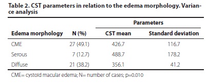
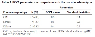
PURPOSE: To correlate the central subfield thickness (CST) measured by CirrusTM SD-OCT with best-corrected visual acuity (BCVA) and structural changes in diabetic macular edema (DME). METHODS: The transversal study evaluated 200 patients with non-proliferative diabetic retinopathy (NPDR) and selected 55 eyes with DME between January, 2010 and April, 2011. Spectral domain OCT was performed in patients with type 2 diabetes and DME. CST and BCVA were correlated with the edema morphology and the ELM (external limiting membrane) integrity. Statistical tests were applied to validate the results. RESULTS: There was no difference between genders in the NPDR classification. 47.3% of the patients showed moderate NPDR. The CST average for male was of 393.58 µm and 434.16 µm for female, with no statistically significant difference. The patients with continuous ELM showed lower CST average (368.73 µm) than those with disrupted ELM (521.43 µm). There was a strong correlation between the macular volume and CST (59.63%), but poor correlation between age and CST (2.9%). Also, there was a significant difference between the average CST and the type of macular edema. Patients with serous detachment showed higher CST average (488.71 µm) than those with cystoid macular edema (CME) and diffuse edema. Patients with severe NPDR showed higher CST average (491.45 µm), if compared to mild and moderate NPDR. Cystoid macular edema was the most common type of edema (49.1%) and showed the worse VA. Patients with disrupted ELM showed worse BCVA. Patients with higher CST showed worse BCVA. There was a significant difference between the CST average of the case group (407.6 ± 113,1 µm) and the control group (diabetic patients without DME: 252 ± 12.5 µm). There was also a significant difference in the BCVA variables and macular volume between case and control groups. CONCLUSION: The study suggests that the CST of diabetic patients with edema is higher than the control group, the increase in CST of diabetic patients with edema leads to worsening of BCVA and macular volume. Continuous ELM showed lower CST average, and the serous detachment showed higher CST average. CirrusTM proved to be an important tool in the DME evaluation.
Diabetic retinopathy; Macular edema; Tomography, optical coherence