Purpose:
The goal of this cross-sectional observational study was to quantify the pattern-shift visual evoked potentials (VEP) and the thickness as well as the volume of retinal layers using optical coherence tomography (OCT) across a cohort of Parkinson's disease (PD) patients and age-matched controls.
Methods:
Forty-three PD patients and 38 controls were enrolled. All participants underwent a detailed neurological and ophthalmologic evaluation. Idiopathic PD cases were included. Cases with glaucoma or increased intra-ocular pressure were excluded. Patients were assessed by VEP and high-resolution Fourier-domain OCT, which quantified the inner and outer thicknesses of the retinal layers. VEP latencies and the thicknesses of the retinal layers were the main outcome measures.
Results:
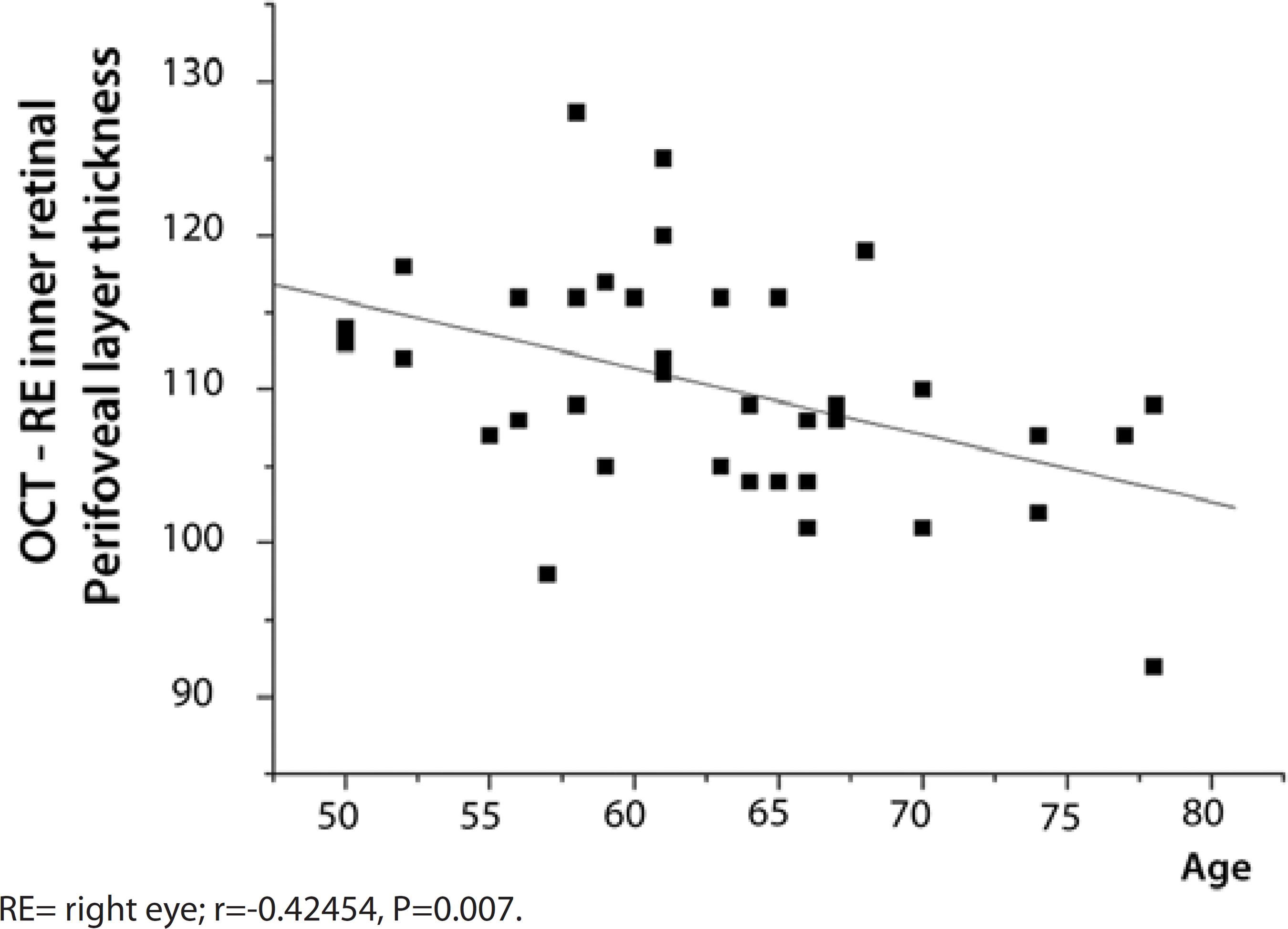
The mean age, with standard deviation (SD), of the PD patients and controls were 63.1 (7.5) and 62.4 (7.2) years, respectively. The patients were predominantly in the initial Hoehn-Yahr (HY) disease stages (34.8% in stage 1 or 1.5, and 55.8 % in stage 2). The VEP latencies and the thicknesses as well as the volumes of the retinal inner and outer layers of the groups were similar. A negative correlation between the retinal thickness and the age was noted in both groups. The thickness of the retinal nerve fibre layer (RNFL) was 102.7 μm in PD patients vs. 104.2 μm in controls.
Conclusions:
The thicknesses of retinal layers, VEP, and RNFL of PD patients were similar to those of the controls. Despite the use of a representative cohort of PD patients and high-resolution OCT in this study, further studies are required to establish the validity of using OCT and VEP measurements as the anatomic and functional biomarkers for the evaluation of retinal and visual pathways in PD patients.
Evoked potentials, visual; Tomography, optical coherence/methods; Parkinson disease