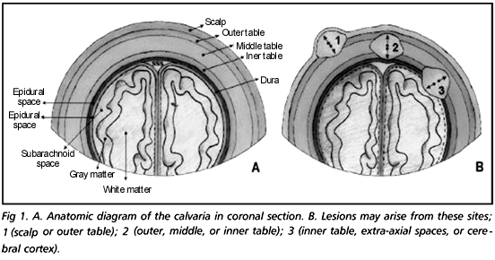
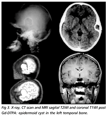
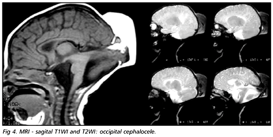
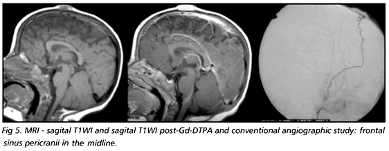
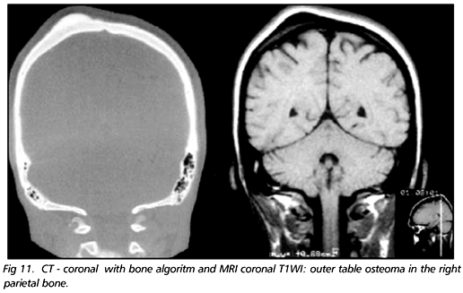
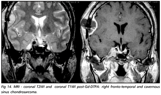
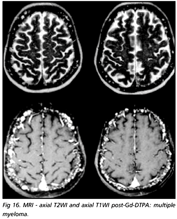
PURPOSE: A variety of diseases affect the calvaria. They may be identified clinically as palpable masses or incidentally in radiologic examinations. There are many diagnostic possibilities, including congenital, neoplastic, inflammatory and traumatic lesions. The purpose of this study is to illustrate the main calvarial lesions through MR imaging, their signal intensity and extension to neighboring sites. METHOD: A retrospective analysis of 81 cases, from November 1996 to July 2001, was conducted. The examinations were performed on a 1.5 T equipment and each one of the cases was pathologically proven. RESULTS: The results were: dermoid cysts [4 cases (5%)], epidermoid cysts [2 cases (2.5%)], cephalocele [14 cases (17.5%)], sinus pericranii [3 cases (3.7%)], leptomeningeal cysts [4 cases (5%)], Langerhans cell histiocytosis [10 cases (12.5%)], lipoma [4 cases (5%)], fibrous dysplasia [13 cases (16.2%)], osteoma [8 cases (10%)], hemangioma [1 case (1.2%)], meningioma [3 cases (3.7%)], chondrosarcoma [5 cases (6.2%)], hemangiosarcoma [1 case (1.2%)], multiple myeloma [3 cases (3.7%)], sarcomatous transformation of Paget disease [1 case (1.3%)], and metastasis [5 cases (6.2%)]. CONCLUSION: MRI identifies bone marrow abnormalities and invasion of adjacent tissues at an early stage. Therefore, it is an essential method when it commes to properly evaluating calvarial lesions.
calvaria; MRI; tumors; congenital lesions; infammatory lesions