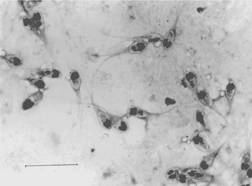
Piauçus (Leporinus macrocephalus), were raised in 300 m² ponds (density of 10 fish/m²) presenting asphyxia signals and daily mortality of 27 fishes. Specimens with 8-cm total body length, were collected for necropsy. Mucus of body surface and pieces of organs were collected and examined microscopically, in wet mounts, stained or in histological sections. The smears examination showed the presence of several spores in the secondary lamellae of the gill filaments, identified as Henneguya leporinicola n.sp (Myxozoa: Myxobolidae). Histopatological study showed epithelial hyperplasia and fulfilling of the spaces between the secondary lamellae, congestion and teleangiectasia sinusoidal. It was also observed hyperplasia of the goblet cells and several cysts of parasite with 70.3mum diameter. Such cysts were situated among the secondary lamellae, covered or not by the hyperplasic epithelium. With this diagnostic, three applications of formalin solution 10 ml/m³ were carried out. Fifteen days after that, fish were examined again to ascertain whether the treatment was efficient on disease caused by the protozoa. The tissue alterations present in the gills after the treatment were just a moderate sinusoidal congestion and a slight epithelial hyperplasia on the base of the secondary lamellae.
Myxozoa; Henneguya leporinicola; Leporinus macrocephalus; histopathology; description