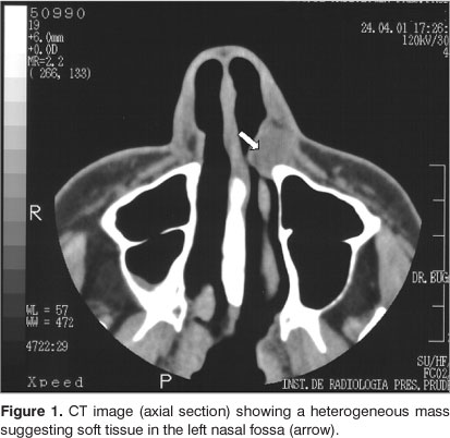
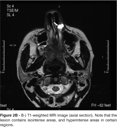
The nasolabial cyst is an uncommon non-odontogenic cyst that develops in the lower region of the nasal ala; its pathogenesis is uncertain. This lesion grows slowly and measures between 1.5 and 3 cm; it is characterized clinically by a floating tumefaction in the nasolabial sulcus, which elevates the upper lip. The diagnosis is based on the clinical findings and, if necessary, image exams. This paper reports a case of a white 48-year-old Brazilian female patient that presented a firm tumor in the left ala of the nose; the clinical features suggested a nasolabial cyst. CT scans revealed an expanding tumor with soft tissue density located in the left ala of the nose. It measured 1.2 cm in diameter and had a clear and well-defined outline; its homogeneous density was about 50 HU. MR images revealed a circular lesion located in soft tissue; T1 and T2 weighted signals were hyperintense, as were images after fat suppression. The diagnosis was a nasolabial cyst, which was confirmed by histopathology after surgery.
nasolabial cyst; maxillary cysts; non-odontogenic cysts