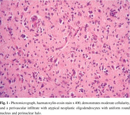
In the last years, new techniques of neuroimages and histopathological methods have been added to the management of cerebral mass lesions in patients with AIDS. Stereotactic biopsy is necessary when after 14 days of empirical treatment for Toxoplasma gondii encephalitis there is no clinical or neuroradiologic improvement. We report a woman with AIDS who developed a single focal brain lesion on the right frontal lobe. She presented a long history of headache and seizures. After two weeks of empirical treatment for toxoplasma encephalitis without response, a magnetic resonance image with spectroscopy was performed and showed a tumoral pattern with a choline peak, diminished of N-acetyl-aspartate and presence of lactate. A stereotactic biopsy was performed. Histopathological diagnosis was a diffuse oligodendroglioma type A. A microsurgical resection of the tumor was carried out and antiretroviral treatment was started. To date she is in good clinical condition, with undetectable plasma viral load and CD4 T cell count > 200 cell/uL.
Focal brain mass; Oligodendroglioma; Acquired immunodeficiency syndrome; AIDS


