ABSTRACT
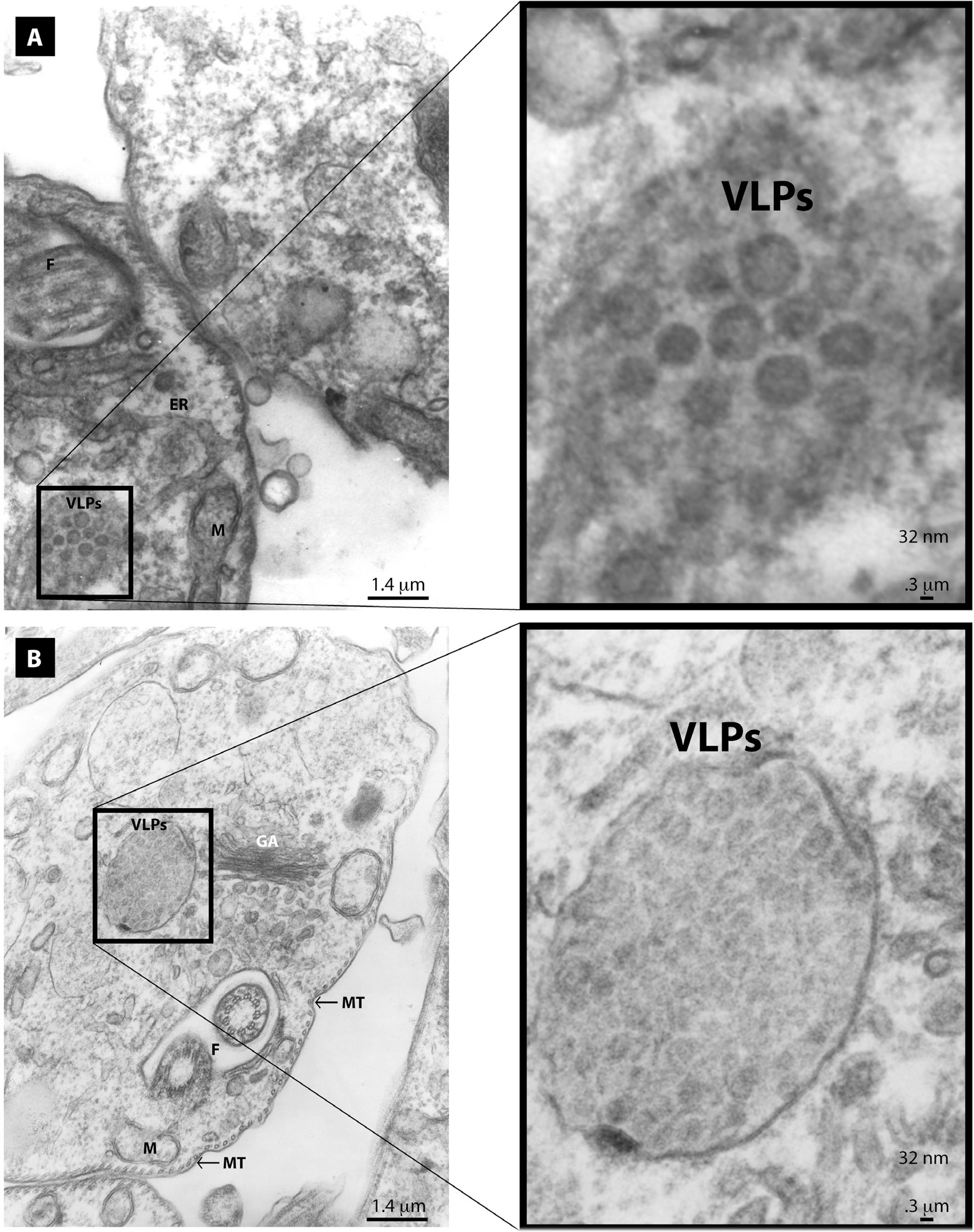
Electron microscopy is routinely used to identify viral infections in protozoan parasites. These viruses have been described as non-enveloped and icosahedral structures with a diameter of 30-60 nm. Most of them are classified within the non-segmented dsRNA Totiviridae family. We observed virus-like particles (VLPs) through transmission electron microscopy in the cytoplasm of Trypanosoma cruzi epimastigotes grown in cultures. Clusters of electrodense enveloped VLPs having a diameter of 48 nm were also observed. These clusters appear to have been released from distended Golgi cisternae. Furthermore, a paracrystalline array of electrodense, non-enveloped VLPs (with a diameter of 32 nm) were found in distended Golgi cisternae or as smaller clusters at a distance from the RE or Golgi. We cannot rule out that the 48 nm enveloped VLPs belong to the ssRNA Flaviviridae family because they are within its size range. The localization of enveloped VLPs is consistent with the replication strategy of these viruses that transit through the Golgi to be released at the cell surface. Due to the size and shape of the 32 nm non-enveloped VLPs, we propose that they belong to the dsRNA Totiviridae family. This is the first description of cytoplasmic enveloped and non-enveloped VLPs in T. cruzi epimastigotes.
Trypanosoma cruzi; Arthropod-borne viruses; Protozoan infections; Flaviviridae; Totiviridae