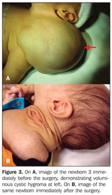
OBJECTIVE: To evaluate three cases of cervical lymphangioma with magnetic resonance imaging and correlating with sonographic findings. MATERIALS AND METHODS: Three pregnant women between the 24th and 35th gestational weeks, with sonographic findings suggestive of fetal cystic hygroma, were submitted to magnetic resonance and subsequently to a new ultrasonography for correlation of imaging findings. Tumors size, location, content and extent were evaluated both at magnetic resonance imaging and ultrasonography. RESULTS: Findings regarding tumor location, size and content were similar for both methods. All the lesions were found in the posterior and lateral cervical space. As regards the tumors content, two of the lesions were predominantly cystic, with thin septations, and the other was heterogeneous. Lesions extent and adjacent structures invasion were better characterized by magnetic resonance imaging, with appropriate demonstration of invasion of the pinna in one case and invasion of the superior mediastinum in another. CONCLUSION: Fetal magnetic resonance imaging can be a useful adjuvant to obstetric ultrasonography in cases of lymphangioma because of its higher accuracy in the determination of these tumors extent and adjacent structures invasion, allowing a better postnatal surgical planning.
Fetus; Lymphangioma; Cystic hygroma; Ultrasonography; Fetal magnetic resonance imaging