Abstract
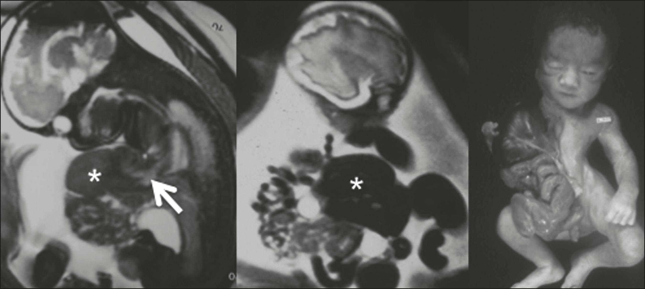
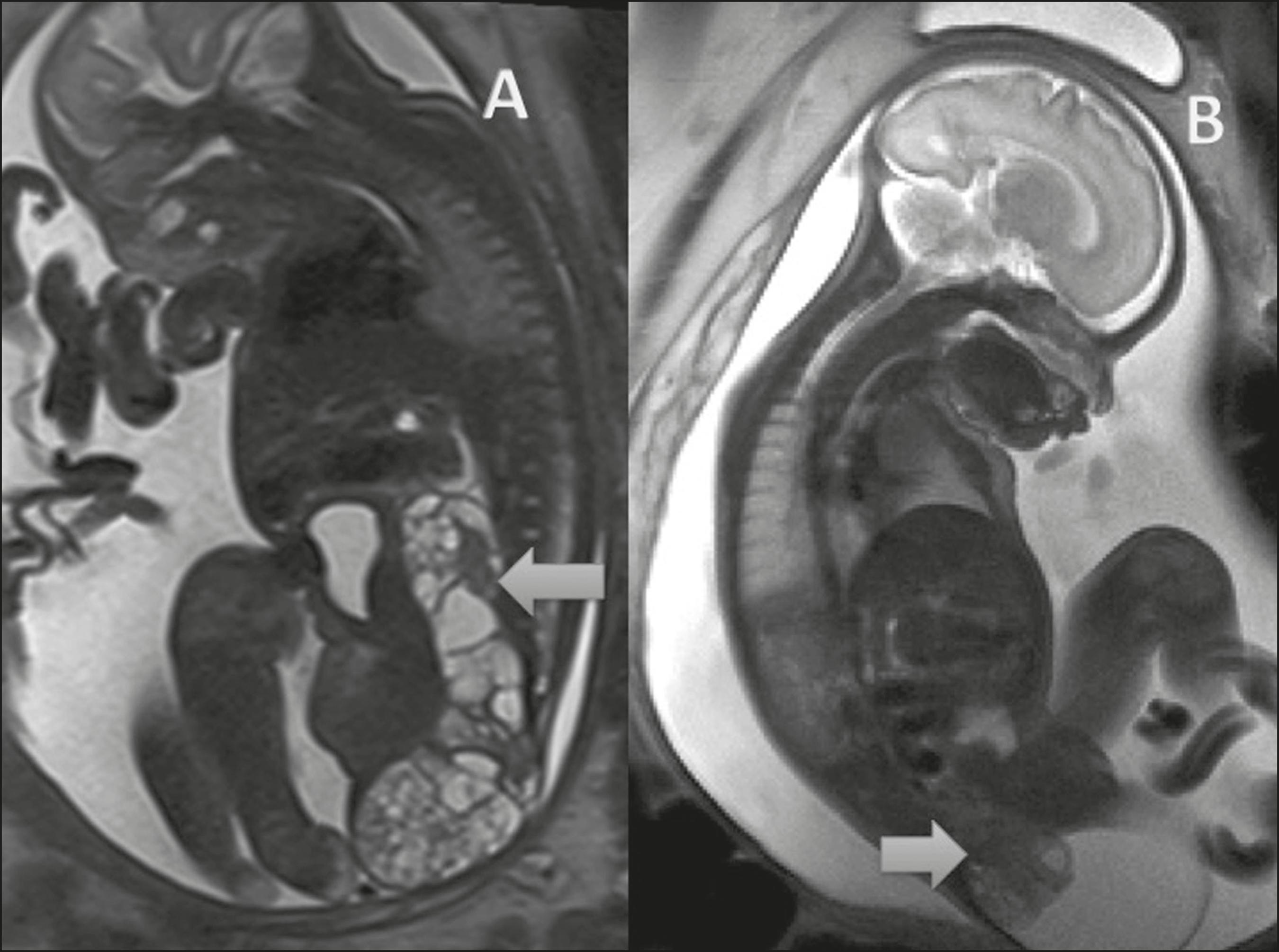
Although ultrasound is still the gold standard for the assessment of fetal malformations, magnetic resonance imaging (MRI) has gained great prominence in recent years. In situations in which ultrasound has low sensitivity, such as maternal obesity, abdominal scarring, and oligohydramnios, MRI has proven to be a safe and accurate method. Regarding fetal abdominal wall defects, MRI appears to be widely used in the prognostic assessment of gastroschisis with intestinal atresia or of complications of omphalocele, allowing better perinatal management and parental counseling. In addition, MRI allows the assessment of local invasion of fetal abdominal tumors, with significant prognostic value for the postnatal period. In this article, we review the main MRI findings in the evaluation of fetal abdominal wall defects and tumors.
Keywords:
Fetus; Congenital abnormalities/diagnostic imaging; Abdomen/diagnostic imaging; Magnetic resonance imaging