ABSTRACT:
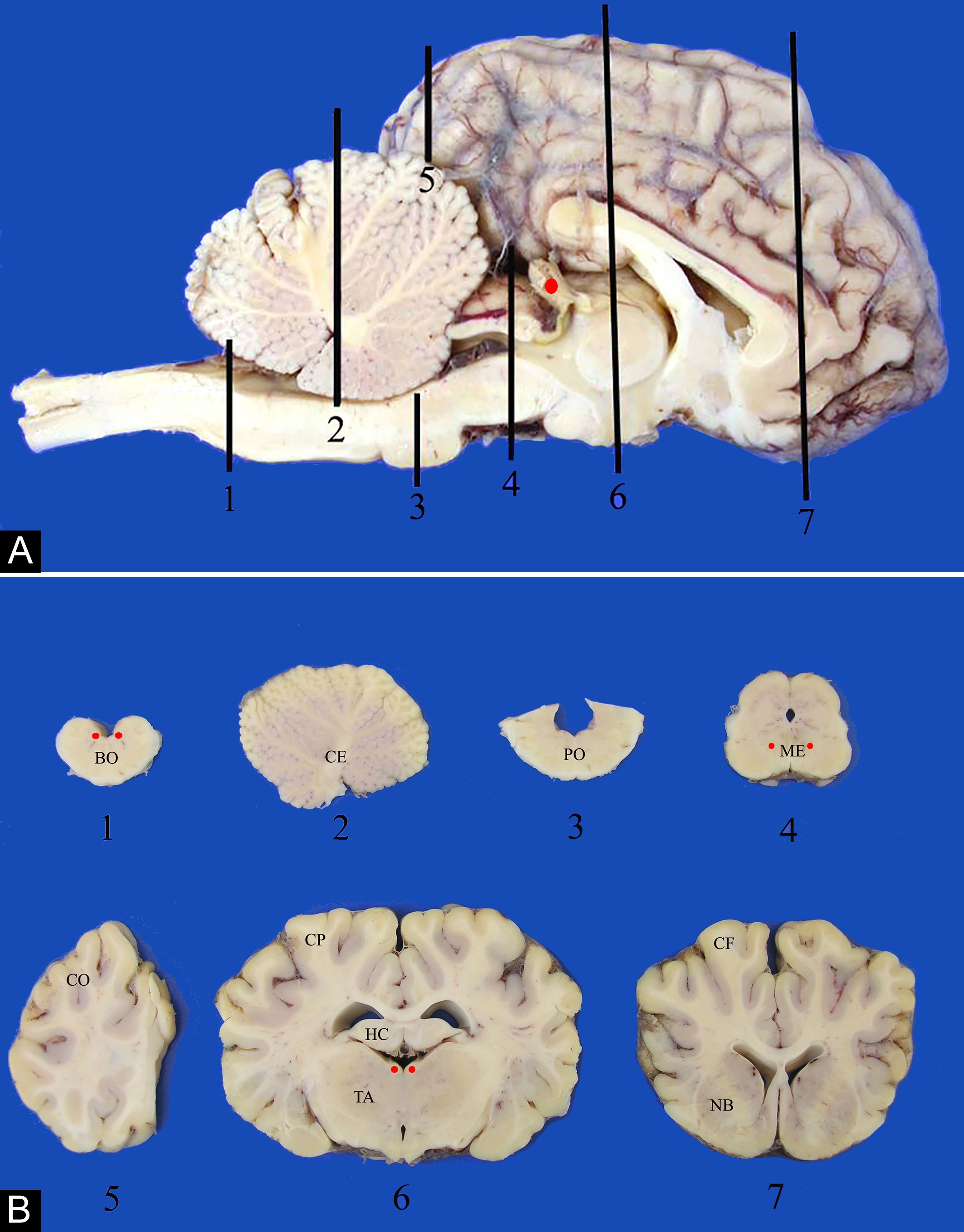
This study stems from the findings during the gross and histopathological exam of 3,338 cattle brains as part of the bovine spongiform encephalopathy (BSE) active surveillance program of the Brazilian Ministry of Agriculture, Livestock, and Supply from 2001 to 2005. The work was carried out in the Veterinary Pathology Laboratory of the Federal University of Santa Maria which at the time (2001-2007) was the national reference laboratory for the diagnosis of BSE and other transmissible spongiform encephalopathies. Both gross and histopathological aspects are described. Several gross aspects were annotated: anatomic normal structures not commonly recognized (non-lesions), lesions of no clinical significance, postmortem changes and artifacts; all these can amount to important pitfalls that distract the pathologist during the routine gross examination of the central nervous system (CNS). Accordingly, equivalent pitfalls were described in the histological examination. Non-lesions observed were the pineal body, embryo remnants such as the external germinal layer of the cerebellum, subependymal plates, and clusters of neuroblasts in the basal ganglia; or circumventricular structures such as area postrema, subcomisural organ, and melanosis in the leptomeninges and vessel walls. Lesions with little or no clinical importance included age-related changes as lipofuscin, hemosiderin, mineralization and hyalinization of vessel walls within the brain and meninges. Corpora amylacea and corpora arenacea were detected respectively in astrocyte processes and the pineal body. Cytoplasmic neuronal vacuolization was observed in the red nucleus and habenular nucleus. Sarcocystis sp. without a correspondent inflammatory reaction was rarely observed. Included within findings with no clinical manifestation were axonal spheroids and perivascular mononuclear cuffings. Changes in the CNS due to killing, sampling and fixation methods can obscure or distract from the more critical lesions. The ones related to the process of killing included hemorrhages caused in cattle destroyed by a captive bolt. Artifacts related to sampling and handling of CNS tissue consisted of inclusion of bone sand in the neural tissue from sawing the calvarium; dark neurons produced by excessive handling of the brain, and micro-organisms that contaminated the tissues during sampling or histological processing. Postmortem autolytic or putrefactive changes observed included vacuolar changes in the myelin sheath, clear halos surrounding neurons and oligodendrocytes, clusters of putrefaction bacilli within vessels or dispersed throughout the brain tissue associated or not to clear halos. One interesting, and somewhat frequent, postmortem autolytic change found in the bovine brain was the partial dissolution of the granule cell layer (GCL) of the cerebellum, also referred to as conglutination of the GCL or as the French denomination “état glace”. Due to the shortage of comprehensive publications in the subject, this review is intended to address the main pitfalls that can be observed in the brain of cattle hoping to help other pathologists avoiding misinterpret them.
INDEX TERMS:
Postmortem findings; histopathology; pitfalls; cattle; brain; Brazil; central nervous system; artifacts; histology; neuropathology
 Thumbnail
 Thumbnail
 Thumbnail
 Thumbnail
 Thumbnail