
ABSTRACT:
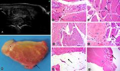
Although ultrasound (US) is a routine diagnostic modality, it still presents limitations for the diagnosis of lesions such as those in the proximal insertion of the suspensory ligament (PISL) because of its composition, which includes muscle fibers and adipose tissue interspersed with the ligament fibers. The objective of the present study was to describe the ultrasonographic, macroscopic and histological aspects of the PISL of thoracic limbs (TL) and pelvic limbs (PL) in Crioulo horses (CH). We selected 34 specimens of TL (right and left) and 10 specimens of PL of horses with a mean age of 5.7 years, from a private clinic or sent to the Department of Veterinary Pathology of UFSM, which died from different causes. The animals had no previous history of lameness in selected limbs associated with PISL injuries. The 34 specimens of PISL of TL were divided into CH (n=25) and Thoroughbred horses (TBH) (n=9), which composed the control group, and 11 specimens of PISL of PL were divided into CH (n=8) and TBH (n=3), which also served as control. The US examination was performed in the PISL using a Sonosite Edge device, 5-10 MHz linear transducer, with cross-sectional and longitudinal palmaromedial and palmarolateral images of the proximal surface of metacarpus III, II and IV (MCIII/MCII/MCIV). In PL, the evaluation was performed four centimeters below the chestnut in the plantaromedial aspect of metatarsus III and II (MTIII/MTII). PISL lobulated shape and size were compared with those of the contralateral limb, as well as the regularity of the palmar bone surface of MC III, II and IV. Subsequently, dissection of the PISL lobes was performed, as well as its macroscopic evaluation, which preceded the histological processing of the samples. In specimens of the CH breed, PISL showed echogenicity varying from peripheral dorsal hyperechogenic zones that merge into echogenic and hypoechogenic zones, where lobulation occurs. In the samples from the TBH group, PISL was also lobulated, but with differences in the echogenicity pattern such as diffuse hypoechogenicity and echogenicity. Macroscopically, CH samples presented a large amount of adipose tissue that corresponds to the dorsal peripheral zone of PISL, which ends in the connective tissue that delimits the ligamentous lobes. On a macroscopic cross-section of PISL, muscle fibers in red are mixed with white ligament fibers in the center of the ligament. This macroscopic finding was not observed in TBH samples, in which muscle fibers overlap ligament fibers throughout the ligament extension and a small amount of fat is present in the dorsal periphery of the ligament. PISL of PL had a triangular shape with echogenicity characteristics very similar to those observed in TL. In ultrasonographic, macroscopic and histological evaluation, PISL samples of TL and PL in CH showed a larger amount of peripheral dorsal adipose tissue, as well as a larger number of merged ligament and muscle fibers compared with those in TBH.
INDEX TERMS:
Ultrasonography; macroscopic assessment; histology; proximal insertion; suspensory ligament; Crioulo horses; ultrasound; equine; horses
 Thumbnail
 Thumbnail
 Thumbnail
 Thumbnail


