ABSTRACT
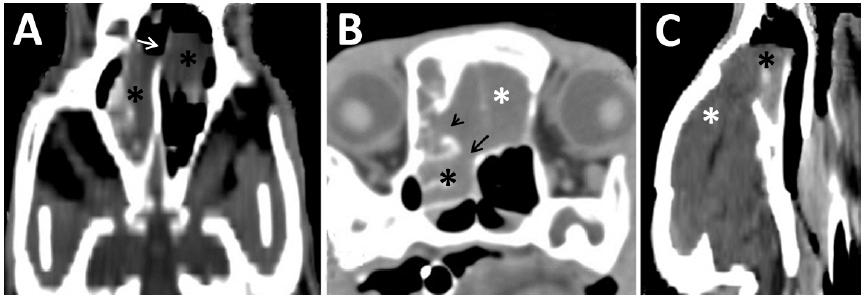
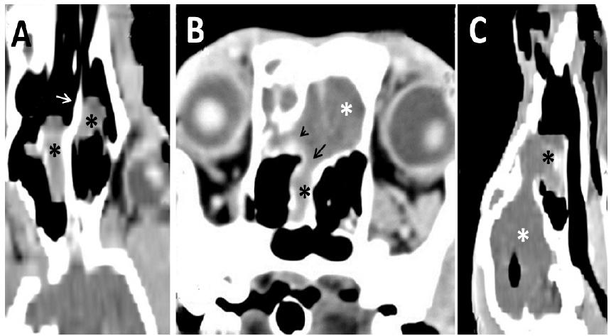
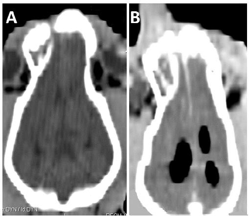
Pneumocephalus is defined as the presence of air in any of the intracranial compartments. Its most frequent causes are trauma and cranial surgery. Clinical signs occur as a result of increased intracranial pressure and vary with the location and extent of the lesion. A case involving a seven-month-old female Saint Bernard, who suffered cranial trauma caused by a bite to the face at ten days of age and had presented with seizures and localized pain four months previously is reported. A computed tomography scan of the skull revealed a nasocephalic mass with low contrast enhancement, bone lysis, and hypodensity (-940 Hounsfield units) of the lateral and third ventricles, indicating intraventricular pneumocephalus. During surgery, a fragment of the mass was collected for histopathological examination, which demonstrated the presence of multifocal areas of necrosis. The computed tomography (CT) is a reliable method for the characterization of intracranial lesions and diagnosis of pneumocephalus, whose occurrence must be considered in pathological processes in which there is increased intracranial pressure and in patients undergoing certain surgical procedures and anesthetic specific, and CT is indicated as a monitoring tool for these patients.
Keywords:
dog; computed tomography; intracerebral aerocele; pneumoventricle