

ABSTRACT
Background:
Gastrointestinal neuroendocrine tumors are rare, usually presented as subepithelial or polypoid tumors. Accurate diagnosis and indication of the type of resection are still challenging.
Aim:
To determine the effectiveness of echoendoscopy in determining the depth of the lesions (T) identified by endoscopy in order to evaluate surgical and/or endoscopic indication, and to evaluate the results of endoscopic removal in the medium term.
Methods:
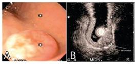
Twenty-seven patients were included, all of whom underwent echoendoscopy for TN tumor staging and the evaluation of possible endoscopic resection. The parameters were: lesion size, origin layer, depth of involvement and identified perilesional adenopathies. The inclusion criteria for endoscopic resection were: 1) high surgical risk; 2) those with NET <2 cm; 3) absence of impairment of the muscle itself; and 4) absence of perilesional adenopathies in echoendoscopy and in others without distant metastases. Exclusion criteria were TNE> 2 cm; those with infiltration of the muscle itself; with perilesional adenopathies and distant metastases. The techniques used were: resection with polypectomy loop; mucosectomy with saline injection; and mucosectomy after ligation with an elastic band. The anatomopathological study of the specimens included evaluation of the margins and immunohistochemistry (chromogranin, synaptophysin and Ki 67) to characterize the tumor. Follow-up was done at 1, 6 and 12 months.
Results:
Resections with polypectomy loop were performed in 15 patients; mucosectomy in five; mucosectomy and ligation with elastic band in three and the remaining four were referred for surgery. The anatomopathological specimens and immunohistochemical analyzes showed positive chromogranin and synaptophysin, while Ki 67 was less than 5% among all cases. The medium-term follow-up revealed three recurrences. The average size of tumors in the stomach was 7.6 mm and in the duodenum 7.2 mm. Well-demarcated, hypoechoic, homogeneous lesions occurred in 75%; mucous layer in 80%; and the deep and submucosal mucosa in 70%.
Conclusions:
Echoendoscopy proved to be a good method for the study of subepithelial lesions, being able to identify the layer affected by the neoplasm, degree of invasion, echogenicity, heterogeneity, size of the lesion and perilesional lymph node involvement and better indicate the treatment option.
HEADING:
Endosonography; Carcinoid Tumor; Carcinoma, Neuroendocrine
 Thumbnail
 Thumbnail


