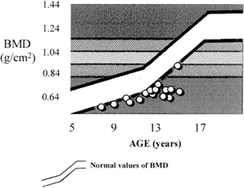
Twenty eight children and adolescents from 7 to 19 years of age, suffering from hepatosplenic mansonic schistosomiasis and bleeding esophageal varices were evaluated for bone mineral density (BMD), before undergoing medical and surgical treatment. The surgical protocol was splenectomy, autoimplantation of spleen tissue into a pouch of the greater omentum and ligature of the left gastric vein. Twenty one patients were evaluated after a follow up from two to nine years post surgical treatment. The BMD was measured at the lumbar spine (L2 - L4) through the dual energy absorptionmetry X-ray (DEXA), using a LUNAR DPX-L densitometer. Preoperatively, all patients showed deficit of the BMD varying from 1 to 7.07 standard deviations (Mean <FONT FACE="Symbol">±</FONT> SEM - 2.64 <FONT FACE="Symbol">±</FONT> 0.28), considering the mean line of the control curve for healthy children accepted as normal. The BMD deficit was more evident among the females than the males. After treatment there was a significant increment (<FONT FACE="Symbol">C</FONT>2 = 9.19 - p =0.01) of the BMD and 29% of the patients (six out of twenty one) were considered without bone mineral deficit. It was concluded that the patients included in this series, who suffer from hepatosplenic mansonic schistosomiasis, showed an important BMD deficit, specially among the females which has had a significant improvement after medical and surgical treatment.
Schistosomiasis mansoni; Hypertension; Splenectomy; Bone density


